 Movie
Movie Controller

[English] 日本語
 Yorodumi
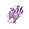
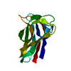
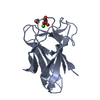
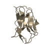
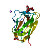
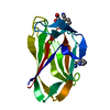
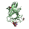
- PDB-1uxx: CBM6ct from Clostridium thermocellum in complex with xylopentaose -
+ Open data

- Basic information
| Entry | Database: PDB / ID: 1uxx | |||||||||
|---|---|---|---|---|---|---|---|---|---|---|
| Title | CBM6ct from Clostridium thermocellum in complex with xylopentaose | |||||||||
 Components Components | XYLANASE U | |||||||||
 Keywords Keywords | CARBOHYDRATE BINDING MODULE / CBM6 / XYLOPENTAOSE BINDING / XYLAN DEGRADATION | |||||||||
| Function / homology |  Function and homology information Function and homology informationhydrolase activity, acting on carbon-nitrogen (but not peptide) bonds / endo-1,4-beta-xylanase / endo-1,4-beta-xylanase activity / xylan catabolic process / carbohydrate binding / metal ion binding Similarity search - Function | |||||||||
| Biological species |  CLOSTRIDIUM THERMOCELLUM (bacteria) CLOSTRIDIUM THERMOCELLUM (bacteria) | |||||||||
| Method |  X-RAY DIFFRACTION / SYNCHROTRON / MOLECULAR REPLACEMENT / Resolution: 1.6 Å X-RAY DIFFRACTION / SYNCHROTRON / MOLECULAR REPLACEMENT / Resolution: 1.6 Å | |||||||||
 Authors Authors | Czjzek, M. / Pires, V.M.R. / Henshaw, J. / Prates, J.A.M. / Henrissat, D.B.B. / Gilbert, H.J. | |||||||||
 Citation Citation | Journal: J.Biol.Chem. / Year: 2004 Title: The Crystal Structure of the Family 6 Carbohydrate Binding Module from Cellvibrio Mixtus Endoglucanase 5A in Complex with Oligosaccharides Reveals Two Distinct Binding Sites with Different Ligand Specificities Authors: Pires, V.M.R. / Henshaw, J. / Prates, J.A.M. / Bolam, D. / Ferreira, L.M.A. / Fontes, C.M.G.A. / Henrissat, B. / Planas, A. / Gilbert, H.J. / Czjzek, M. | |||||||||
| History |
|
- Structure visualization
| Structure viewer | Molecule: MolmilJmol/JSmol |
|---|
- Downloads & links
-Download
| PDBx/mmCIF format | 1uxx.cif.gz | 42.4 KB | Display | PDBx/mmCIF format |
|---|---|---|---|---|
| PDB format | pdb1uxx.ent.gz | 27.6 KB | Display | PDB format |
| PDBx/mmJSON format | 1uxx.json.gz | Tree view | PDBx/mmJSON format | |
| Others |  Other downloads Other downloads |
-Validation report
| Arichive directory | https://data.pdbj.org/pub/pdb/validation_reports/ux/1uxxftp://data.pdbj.org/pub/pdb/validation_reports/ux/1uxx | HTTPS FTP |
|---|
-Related structure data
| Related structure data | 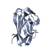 1uxzC 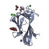 1uy0C 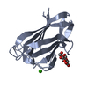 1uyxC 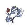 1uyyC 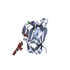 1uyzC 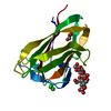 1uz0C 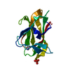 1gmmS S: Starting model for refinement C: citing same article ( |
|---|---|
| Similar structure data |


-Links
 PDBj
PDBj

- Assembly
| Deposited unit | 
| ||||||||
|---|---|---|---|---|---|---|---|---|---|
| 1 |
| ||||||||
| Unit cell |
|
-Components
| #1: Protein | Mass: 14115.347 Da / Num. of mol.: 1 / Fragment: CARBOHYDRATE BINDING MODULE, RESIDUES 248-380 Source method: isolated from a genetically manipulated source Source: (gene. exp.) CLOSTRIDIUM THERMOCELLUM (bacteria) / Production host: |
|---|---|
| #2: Polysaccharide | beta-D-xylopyranose-(1-4)-beta-D-xylopyranose-(1-4)-beta-D-xylopyranose-(1-4)-beta-D-xylopyranose- ...beta-D-xylopyranose-(1-4)-beta-D-xylopyranose-(1-4)-beta-D-xylopyranose-(1-4)-beta-D-xylopyranose-(1-4)-beta-D-xylopyranose Source method: isolated from a genetically manipulated source |
| #3: Chemical | ChemComp-CA /   Mass: 40.078 Da / Num. of mol.: 1 / Source method: obtained synthetically / Formula: Ca Mass: 40.078 Da / Num. of mol.: 1 / Source method: obtained synthetically / Formula: Ca |
| #4: Water | ChemComp-HOH /  Mass: 18.015 Da / Num. of mol.: 108 / Source method: isolated from a natural source / Formula: H2O Mass: 18.015 Da / Num. of mol.: 108 / Source method: isolated from a natural source / Formula: H2O |
| Compound details | CATALYSES THE ENDOHYDROLYSIS OF 1,4-BETA-D-XYLOSIDIC LINKAGES IN XYLANS. A MEMBER OF THE XYLAN ...CATALYSES THE ENDOHYDROL |
| Sequence details | CBM6 IS A FRAGMENT OF THIS SEQUENCE |
-Experimental details
-Experiment
| Experiment | Method: X-RAY DIFFRACTION / Number of used crystals: 1 |
|---|
- Sample preparation
| Crystal | Density Matthews: 1.9 Å3/Da / Density % sol: 35 % | ||||||||||||||||||||||||
|---|---|---|---|---|---|---|---|---|---|---|---|---|---|---|---|---|---|---|---|---|---|---|---|---|---|
| Crystal grow | pH: 4.6 Details: 20% PEG 1000, 0.2 M SODIUM ACETATE, 0.1 M ACETATE BUFFER PH4.6, pH 4.60 | ||||||||||||||||||||||||
| Crystal grow | *PLUS Method: vapor diffusion, hanging drop | ||||||||||||||||||||||||
| Components of the solutions | *PLUS
|
-Data collection
| Diffraction | Mean temperature: 100 K |
|---|---|
| Diffraction source | Source: SYNCHROTRON / Site: ESRF  / Beamline: ID14-1 / Wavelength: 0.934 / Beamline: ID14-1 / Wavelength: 0.934 |
| Detector | Date: Mar 15, 2003 |
| Radiation | Protocol: SINGLE WAVELENGTH / Monochromatic (M) / Laue (L): M / Scattering type: x-ray |
| Radiation wavelength | Wavelength: 0.934 Å / Relative weight: 1 |
| Reflection | Resolution: 1.6→52 Å / Num. obs: 13033 / % possible obs: 99.5 % / Redundancy: 3.4 % / Rmerge(I) obs: 0.03 / Net I/σ(I): 16 |
| Reflection shell | Resolution: 1.6→1.66 Å / Redundancy: 3.4 % / Rmerge(I) obs: 0.094 / Mean I/σ(I) obs: 9.1 / % possible all: 99.9 |
| Reflection | *PLUS Highest resolution: 1.6 Å / Redundancy: 3.4 % / Rmerge(I) obs: 0.03 |
| Reflection shell | *PLUS % possible obs: 99.9 % / Redundancy: 3.4 % / Rmerge(I) obs: 0.094 / Mean I/σ(I) obs: 9.1 |
- Processing
| Software |
| ||||||||||||||||||||||||||||||||||||||||||||||||||||||||||||||||||||||||||||||||||||||||||||||||||||||||||||||||||||||||||||||||||||||||||||||||||||||||||||||||||||||||||||||||||||||
|---|---|---|---|---|---|---|---|---|---|---|---|---|---|---|---|---|---|---|---|---|---|---|---|---|---|---|---|---|---|---|---|---|---|---|---|---|---|---|---|---|---|---|---|---|---|---|---|---|---|---|---|---|---|---|---|---|---|---|---|---|---|---|---|---|---|---|---|---|---|---|---|---|---|---|---|---|---|---|---|---|---|---|---|---|---|---|---|---|---|---|---|---|---|---|---|---|---|---|---|---|---|---|---|---|---|---|---|---|---|---|---|---|---|---|---|---|---|---|---|---|---|---|---|---|---|---|---|---|---|---|---|---|---|---|---|---|---|---|---|---|---|---|---|---|---|---|---|---|---|---|---|---|---|---|---|---|---|---|---|---|---|---|---|---|---|---|---|---|---|---|---|---|---|---|---|---|---|---|---|---|---|---|---|
| Refinement | Method to determine structure: MOLECULAR REPLACEMENT Starting model: PDB ENTRY 1GMM Resolution: 1.6→51.99 Å / Cor.coef. Fo:Fc: 0.951 / Cor.coef. Fo:Fc free: 0.922 / SU B: 1.71 / SU ML: 0.062 / Cross valid method: THROUGHOUT / ESU R: 0.108 / ESU R Free: 0.107 / Stereochemistry target values: MAXIMUM LIKELIHOOD / Details: HYDROGENS HAVE BEEN ADDED IN THE RIDING POSITIONS
| ||||||||||||||||||||||||||||||||||||||||||||||||||||||||||||||||||||||||||||||||||||||||||||||||||||||||||||||||||||||||||||||||||||||||||||||||||||||||||||||||||||||||||||||||||||||
| Solvent computation | Ion probe radii: 0.8 Å / Shrinkage radii: 0.8 Å / VDW probe radii: 1.4 Å / Solvent model: BABINET MODEL WITH MASK | ||||||||||||||||||||||||||||||||||||||||||||||||||||||||||||||||||||||||||||||||||||||||||||||||||||||||||||||||||||||||||||||||||||||||||||||||||||||||||||||||||||||||||||||||||||||
| Displacement parameters | Biso mean: 13.52 Å2
| ||||||||||||||||||||||||||||||||||||||||||||||||||||||||||||||||||||||||||||||||||||||||||||||||||||||||||||||||||||||||||||||||||||||||||||||||||||||||||||||||||||||||||||||||||||||
| Refinement step | Cycle: LAST / Resolution: 1.6→51.99 Å
| ||||||||||||||||||||||||||||||||||||||||||||||||||||||||||||||||||||||||||||||||||||||||||||||||||||||||||||||||||||||||||||||||||||||||||||||||||||||||||||||||||||||||||||||||||||||
| Refine LS restraints |
|