 Movie
Movie Controller

[English] 日本語
 Yorodumi
- PDB-1usm: DCOH, A BIFUNCTIONAL PROTEIN-BINDING TRANSCRIPTIONAL COACTIVATOR,... -
+ Open data

- Basic information
| Entry | Database: PDB / ID: 1usm | ||||||
|---|---|---|---|---|---|---|---|
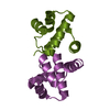
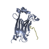
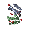
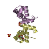
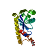
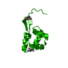
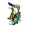
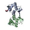
| Title | DCOH, A BIFUNCTIONAL PROTEIN-BINDING TRANSCRIPTIONAL COACTIVATOR, PRO9LEU MUTANT | ||||||
 Components Components | HEPATOCYTE NUCLEAR FACTOR 1-ALPHA | ||||||
 Keywords Keywords | TRANSCRIPTIONAL STIMULATOR / DIMERIZATION COFACTOR / DEHYDRATASE / 4A-CARBINOLAMINE DEHYDRATASE / TRANSREGULATOR OF HOMEODOMAIN PROTEINS / RIKEN STRUCTURAL GENOMICS/PROTEOMICS INITIATIVE / RSGI / STRUCTURAL GENOMICS | ||||||
| Function / homology |  Function and homology information Function and homology information4a-hydroxytetrahydrobiopterin dehydratase / 4-alpha-hydroxytetrahydrobiopterin dehydratase activity / tetrahydrobiopterin biosynthetic process Similarity search - Function | ||||||
| Biological species |   THERMUS THERMOPHILUS (bacteria) THERMUS THERMOPHILUS (bacteria) | ||||||
| Method |  X-RAY DIFFRACTION / SYNCHROTRON / MOLECULAR REPLACEMENT / Resolution: 1.2 Å X-RAY DIFFRACTION / SYNCHROTRON / MOLECULAR REPLACEMENT / Resolution: 1.2 Å | ||||||
 Authors Authors | Tahirov, T.H. / Inagaki, E. | ||||||
 Citation Citation | Journal: To be Published Title: Crystal Structure of Transcriptional Coactivator DCoH from Thermus Thermophilus Authors: Tahirov, T.H. / Inagaki, E. | ||||||
| History |
|
- Structure visualization
| Structure viewer | Molecule: MolmilJmol/JSmol |
|---|
- Downloads & links
-Download
| PDBx/mmCIF format | 1usm.cif.gz | 31.7 KB | Display | PDBx/mmCIF format |
|---|---|---|---|---|
| PDB format | pdb1usm.ent.gz | 21.2 KB | Display | PDB format |
| PDBx/mmJSON format | 1usm.json.gz | Tree view | PDBx/mmJSON format | |
| Others |  Other downloads Other downloads |
-Validation report
| Summary document | 1usm_validation.pdf.gz | 409.8 KB | Display | wwPDB validaton report |
|---|---|---|---|---|
| Full document | 1usm_full_validation.pdf.gz | 412 KB | Display | |
| Data in XML | 1usm_validation.xml.gz | 7.2 KB | Display | |
| Data in CIF | 1usm_validation.cif.gz | 9.8 KB | Display | |
| Arichive directory | https://data.pdbj.org/pub/pdb/validation_reports/us/1usmftp://data.pdbj.org/pub/pdb/validation_reports/us/1usm | HTTPS FTP |
-Related structure data
| Related structure data |  1usoC 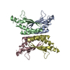 1dcoS S: Starting model for refinement C: citing same article ( |
|---|---|
| Similar structure data |
-Links
 PDBj
PDBj
- Assembly
| Deposited unit | 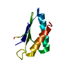
| ||||||||
|---|---|---|---|---|---|---|---|---|---|
| 1 | 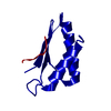
| ||||||||
| Unit cell |
|
-Components
| #1: Protein | Mass: 9508.693 Da / Num. of mol.: 1 Source method: isolated from a genetically manipulated source Details: DIMERIZATION DOMAIN / Source: (gene. exp.) THERMUS THERMOPHILUS (bacteria) / Strain: HB8 / Plasmid: PET11A / Production host: |
|---|---|
| #2: Water | ChemComp-HOH /  Mass: 18.015 Da / Num. of mol.: 147 / Source method: isolated from a natural source / Formula: H2O Mass: 18.015 Da / Num. of mol.: 147 / Source method: isolated from a natural source / Formula: H2O |
| Sequence details | THE RESIDUE PROLINE AT POSITION 9 WAS MUTATED TO LEU |
-Experimental details
-Experiment
| Experiment | Method: X-RAY DIFFRACTION / Number of used crystals: 1 |
|---|
- Sample preparation
| Crystal | Density Matthews: 2.15 Å3/Da / Density % sol: 42.4 % |
|---|---|
| Crystal grow | Method: microbatch / pH: 5.3 Details: MICROBATCH METHOD UNDER OIL BY CRYSTALLIZATION ROBOT TERA. 25.6 MG/ML OF PROTEIN SOLUTION WAS MIXED WITH 3.15M SODIUM FORMATE AND 0.1M SODIUM ACETATE BUFFER, PH 4.0. FINAL PH WAS 5.3. ...Details: MICROBATCH METHOD UNDER OIL BY CRYSTALLIZATION ROBOT TERA. 25.6 MG/ML OF PROTEIN SOLUTION WAS MIXED WITH 3.15M SODIUM FORMATE AND 0.1M SODIUM ACETATE BUFFER, PH 4.0. FINAL PH WAS 5.3. CRYOPROTECTANT CONTENT WAS: 25% GLYCEROL, 2.0M SODIUM FORMATE, AND 0.6M SODIUM ACETATE BUFFER, PH 4.0. |
-Data collection
| Diffraction | Mean temperature: 100 K |
|---|---|
| Diffraction source | Source: SYNCHROTRON / Site: SPring-8  / Beamline: BL26B1 / Wavelength: 0.8 / Beamline: BL26B1 / Wavelength: 0.8 |
| Detector | Type: RIGAKU IMAGE PLATE RAXIS-V / Detector: IMAGE PLATE / Date: Feb 15, 2003 |
| Radiation | Protocol: SINGLE WAVELENGTH / Monochromatic (M) / Laue (L): M / Scattering type: x-ray |
| Radiation wavelength | Wavelength: 0.8 Å / Relative weight: 1 |
| Reflection | Resolution: 1.2→20 Å / Num. obs: 30991 / % possible obs: 98.7 % / Observed criterion σ(I): -0.2 / Redundancy: 8.7 % / Biso Wilson estimate: 12.1 Å2 / Rmerge(I) obs: 0.034 / Net I/σ(I): 41.4 |
| Reflection shell | Resolution: 1.2→1.22 Å / Rmerge(I) obs: 0.146 / Mean I/σ(I) obs: 11.7 / % possible all: 99.5 |
- Processing
| Software |
| ||||||||||||||||||||||||||||||||||||||||||||||||||||||||||||||||||||||||||||||||
|---|---|---|---|---|---|---|---|---|---|---|---|---|---|---|---|---|---|---|---|---|---|---|---|---|---|---|---|---|---|---|---|---|---|---|---|---|---|---|---|---|---|---|---|---|---|---|---|---|---|---|---|---|---|---|---|---|---|---|---|---|---|---|---|---|---|---|---|---|---|---|---|---|---|---|---|---|---|---|---|---|---|
| Refinement | Method to determine structure: MOLECULAR REPLACEMENT Starting model: PDB ENTRY 1DCO Resolution: 1.2→18.77 Å / Rfactor Rfree error: 0.006 / Data cutoff high absF: 3515920.09 / Isotropic thermal model: RESTRAINED / Cross valid method: THROUGHOUT / σ(F): 0 Details: RESIDUES 7-9 ARE DISORDERED AND NOT INCLUDED IN THE MODEL
| ||||||||||||||||||||||||||||||||||||||||||||||||||||||||||||||||||||||||||||||||
| Solvent computation | Solvent model: FLAT MODEL / Bsol: 59.4998 Å2 / ksol: 0.393311 e/Å3 | ||||||||||||||||||||||||||||||||||||||||||||||||||||||||||||||||||||||||||||||||
| Displacement parameters | Biso mean: 19.5 Å2
| ||||||||||||||||||||||||||||||||||||||||||||||||||||||||||||||||||||||||||||||||
| Refine analyze |
| ||||||||||||||||||||||||||||||||||||||||||||||||||||||||||||||||||||||||||||||||
| Refinement step | Cycle: LAST / Resolution: 1.2→18.77 Å
| ||||||||||||||||||||||||||||||||||||||||||||||||||||||||||||||||||||||||||||||||
| Refine LS restraints |
| ||||||||||||||||||||||||||||||||||||||||||||||||||||||||||||||||||||||||||||||||
| LS refinement shell | Resolution: 1.2→1.28 Å / Rfactor Rfree error: 0.016 / Total num. of bins used: 6
| ||||||||||||||||||||||||||||||||||||||||||||||||||||||||||||||||||||||||||||||||
| Xplor file |
|
