 Movie
Movie Controller


+ Open data

- Basic information
| Entry | Database: PDB / ID: 1us5 | ||||||
|---|---|---|---|---|---|---|---|
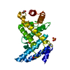
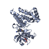
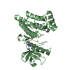
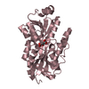
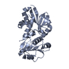
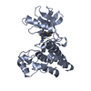
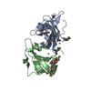
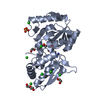
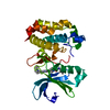
| Title | PUTATIVE GLUR0 LIGAND BINDING CORE WITH L-GLUTAMATE | ||||||
 Components Components | PUTATIVE GLUR0 LIGAND BINDING CORE | ||||||
 Keywords Keywords | RECEPTOR / MEMBRANE PROTEIN / GLUTAMATE RECEPTOR / GLUR0 / L-GLUTAMATE / RIKEN STRUCTURAL GENOMICS/PROTEOMICS INITIATIVE / RSGI / STRUCTURAL GENOMICS | ||||||
| Function / homology | TRAP transporter solute receptor, TAXI family / NMT1-like family / Periplasmic binding protein-like II / D-Maltodextrin-Binding Protein; domain 2 / 3-Layer(aba) Sandwich / Alpha Beta / GLUTAMIC ACID / Glutamate receptor Function and homology information Function and homology information | ||||||
| Biological species |   THERMUS THERMOPHILUS (bacteria) THERMUS THERMOPHILUS (bacteria) | ||||||
| Method |  X-RAY DIFFRACTION / SYNCHROTRON / MOLECULAR REPLACEMENT / Resolution: 1.5 Å X-RAY DIFFRACTION / SYNCHROTRON / MOLECULAR REPLACEMENT / Resolution: 1.5 Å | ||||||
 Authors Authors | Tahirov, T.H. / Inagaki, E. | ||||||
 Citation Citation | Journal: Acta Crystallogr.,Sect.D / Year: 2004 Title: Structure of the Thermus Thermophilus Putative Periplasmic Glutamate/Glutamine-Binding Protein Authors: Takahashi, H. / Inagaki, E. / Kuroishi, C. / Tahirov, T.H. | ||||||
| History |
| ||||||
| Remark 700 | SHEET THE SHEET STRUCTURE OF THIS MOLECULE IS BIFURCATED. IN ORDER TO REPRESENT THIS FEATURE IN ... SHEET THE SHEET STRUCTURE OF THIS MOLECULE IS BIFURCATED. IN ORDER TO REPRESENT THIS FEATURE IN THE SHEET RECORDS BELOW, TWO SHEETS ARE DEFINED. |
- Structure visualization
| Structure viewer | Molecule: MolmilJmol/JSmol |
|---|
- Downloads & links
-Download
| PDBx/mmCIF format | 1us5.cif.gz | 77.4 KB | Display | PDBx/mmCIF format |
|---|---|---|---|---|
| PDB format | pdb1us5.ent.gz | 57.1 KB | Display | PDB format |
| PDBx/mmJSON format | 1us5.json.gz | Tree view | PDBx/mmJSON format | |
| Others |  Other downloads Other downloads |
-Validation report
| Arichive directory | https://data.pdbj.org/pub/pdb/validation_reports/us/1us5ftp://data.pdbj.org/pub/pdb/validation_reports/us/1us5 | HTTPS FTP |
|---|
-Related structure data
| Related structure data | 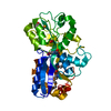 1us4SC S: Starting model for refinement C: citing same article ( |
|---|---|
| Similar structure data |

-Links
 PDBj
PDBj

- Assembly
| Deposited unit | 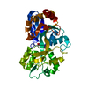
| ||||||||
|---|---|---|---|---|---|---|---|---|---|
| 1 |
| ||||||||
| Unit cell |
|
-Components
| #1: Protein | Mass: 33488.691 Da / Num. of mol.: 1 / Fragment: LIGAND BINDING CORE, RESIDUES 1-314 Source method: isolated from a genetically manipulated source Source: (gene. exp.) THERMUS THERMOPHILUS (bacteria) / Strain: HB8 / Plasmid: PET11A / Production host: |
|---|---|
| #2: Chemical | ChemComp-GLU /   Type: L-peptide linking / Mass: 147.129 Da / Num. of mol.: 1 / Source method: obtained synthetically / Formula: C5H9NO4 Type: L-peptide linking / Mass: 147.129 Da / Num. of mol.: 1 / Source method: obtained synthetically / Formula: C5H9NO4 |
| #3: Chemical | ChemComp-EDO /   Mass: 62.068 Da / Num. of mol.: 1 / Source method: obtained synthetically / Formula: C2H6O2 Mass: 62.068 Da / Num. of mol.: 1 / Source method: obtained synthetically / Formula: C2H6O2 |
| #4: Water | ChemComp-HOH /  Mass: 18.015 Da / Num. of mol.: 288 / Source method: isolated from a natural source / Formula: H2O Mass: 18.015 Da / Num. of mol.: 288 / Source method: isolated from a natural source / Formula: H2O |
-Experimental details
-Experiment
| Experiment | Method: X-RAY DIFFRACTION / Number of used crystals: 1 |
|---|
- Sample preparation
| Crystal | Density Matthews: 2.34 Å3/Da / Density % sol: 47.4 % | ||||||||||||||||||||||||
|---|---|---|---|---|---|---|---|---|---|---|---|---|---|---|---|---|---|---|---|---|---|---|---|---|---|
| Crystal grow | Temperature: 291 K / Method: microbatch / pH: 5 Details: MICROBATCH METHOD UNDER OIL WAS USED. 20 MG/ML OF PROTEIN SOLUTION WAS MIXED WITH 22.5% PEG4000, 1M LITHIUM CHLORIDE AND 0.1M SODIUM CITRATE PH 5. THE CRYSTALLIZATION TEMPERATURE WAS 291 K. ...Details: MICROBATCH METHOD UNDER OIL WAS USED. 20 MG/ML OF PROTEIN SOLUTION WAS MIXED WITH 22.5% PEG4000, 1M LITHIUM CHLORIDE AND 0.1M SODIUM CITRATE PH 5. THE CRYSTALLIZATION TEMPERATURE WAS 291 K. THE CRYSTALS IN FORM OF PARALLELEPIPEDS WERE GROWN TO 0.03X0.03X0.15 MM WITHIN ONE MONTH. CRYOPROTECTANT CONTAINED 25% PEG4000, 18% ETHYLENE GLYCOL, 1M LITHIUM CHLORIDE AND 0.1M SODIUM CITRATE PH 5. | ||||||||||||||||||||||||
| Crystal grow | *PLUS pH: 5 / Method: vapor diffusion, sitting drop | ||||||||||||||||||||||||
| Components of the solutions | *PLUS
|
-Data collection
| Diffraction | Mean temperature: 100 K |
|---|---|
| Diffraction source | Source: SYNCHROTRON / Site: SPring-8  / Beamline: BL26B1 / Wavelength: 1 / Beamline: BL26B1 / Wavelength: 1 |
| Detector | Type: RIGAKU IMAGE PLATE RAXIS-V / Detector: IMAGE PLATE / Date: Feb 15, 2003 |
| Radiation | Protocol: SINGLE WAVELENGTH / Monochromatic (M) / Laue (L): M / Scattering type: x-ray |
| Radiation wavelength | Wavelength: 1 Å / Relative weight: 1 |
| Reflection | Resolution: 1.5→30 Å / Num. obs: 50003 / % possible obs: 97.7 % / Observed criterion σ(I): -0.5 / Redundancy: 4.32 % / Biso Wilson estimate: 13.3 Å2 / Rmerge(I) obs: 0.065 / Net I/σ(I): 20.7 |
| Reflection shell | Resolution: 1.5→1.53 Å / Rmerge(I) obs: 0.362 / Mean I/σ(I) obs: 3.1 / % possible all: 94.7 |
| Reflection | *PLUS Highest resolution: 1.5 Å / Lowest resolution: 20 Å / Num. measured all: 215880 / Rmerge(I) obs: 0.065 |
| Reflection shell | *PLUS Highest resolution: 1.5 Å / % possible obs: 94.7 % / Rmerge(I) obs: 0.362 / Mean I/σ(I) obs: 3.1 |
- Processing
| Software |
| ||||||||||||||||||||||||||||||||||||||||||||||||||||||||||||||||||||||||||||||||
|---|---|---|---|---|---|---|---|---|---|---|---|---|---|---|---|---|---|---|---|---|---|---|---|---|---|---|---|---|---|---|---|---|---|---|---|---|---|---|---|---|---|---|---|---|---|---|---|---|---|---|---|---|---|---|---|---|---|---|---|---|---|---|---|---|---|---|---|---|---|---|---|---|---|---|---|---|---|---|---|---|---|
| Refinement | Method to determine structure: MOLECULAR REPLACEMENT Starting model: PDB ENTRY 1US4 Resolution: 1.5→19.89 Å / Rfactor Rfree error: 0.004 / Data cutoff high absF: 814853.8 / Isotropic thermal model: RESTRAINED / Cross valid method: THROUGHOUT / σ(F): 0
| ||||||||||||||||||||||||||||||||||||||||||||||||||||||||||||||||||||||||||||||||
| Solvent computation | Solvent model: FLAT MODEL / Bsol: 74.25 Å2 / ksol: 0.51198 e/Å3 | ||||||||||||||||||||||||||||||||||||||||||||||||||||||||||||||||||||||||||||||||
| Displacement parameters | Biso mean: 16.9 Å2
| ||||||||||||||||||||||||||||||||||||||||||||||||||||||||||||||||||||||||||||||||
| Refine analyze |
| ||||||||||||||||||||||||||||||||||||||||||||||||||||||||||||||||||||||||||||||||
| Refinement step | Cycle: LAST / Resolution: 1.5→19.89 Å
| ||||||||||||||||||||||||||||||||||||||||||||||||||||||||||||||||||||||||||||||||
| Refine LS restraints |
| ||||||||||||||||||||||||||||||||||||||||||||||||||||||||||||||||||||||||||||||||
| LS refinement shell | Resolution: 1.5→1.59 Å / Rfactor Rfree error: 0.015 / Total num. of bins used: 6
| ||||||||||||||||||||||||||||||||||||||||||||||||||||||||||||||||||||||||||||||||
| Xplor file |
| ||||||||||||||||||||||||||||||||||||||||||||||||||||||||||||||||||||||||||||||||
| Refinement | *PLUS Highest resolution: 1.5 Å / Lowest resolution: 20 Å | ||||||||||||||||||||||||||||||||||||||||||||||||||||||||||||||||||||||||||||||||
| Solvent computation | *PLUS | ||||||||||||||||||||||||||||||||||||||||||||||||||||||||||||||||||||||||||||||||
| Displacement parameters | *PLUS | ||||||||||||||||||||||||||||||||||||||||||||||||||||||||||||||||||||||||||||||||
| Refine LS restraints | *PLUS
| ||||||||||||||||||||||||||||||||||||||||||||||||||||||||||||||||||||||||||||||||
| LS refinement shell | *PLUS Highest resolution: 1.5 Å |
