 Movie
Movie Controller


+ Open data

- Basic information
| Entry | Database: PDB / ID: 1uo9 | ||||||
|---|---|---|---|---|---|---|---|
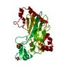
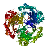
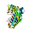
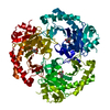
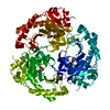
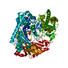
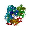
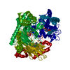
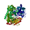
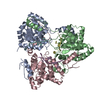
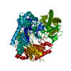
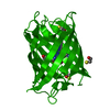
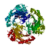
| Title | Deacetoxycephalosporin C synthase complexed with succinate | ||||||
 Components Components | DEACETOXYCEPHALOSPORIN C SYNTHETASE | ||||||
 Keywords Keywords | OXIDOREDUCTASE / ANTIBIOTIC BIOSYNTHESIS / IRON / VITAMIN C | ||||||
| Function / homology |  Function and homology information Function and homology informationdeacetoxycephalosporin-C synthase / deacetoxycephalosporin-C synthase activity / L-ascorbic acid binding / antibiotic biosynthetic process / iron ion binding Similarity search - Function | ||||||
| Biological species |  STREPTOMYCES CLAVULIGERUS (bacteria) STREPTOMYCES CLAVULIGERUS (bacteria) | ||||||
| Method |  X-RAY DIFFRACTION / SYNCHROTRON / MOLECULAR REPLACEMENT / Resolution: 1.5 Å X-RAY DIFFRACTION / SYNCHROTRON / MOLECULAR REPLACEMENT / Resolution: 1.5 Å | ||||||
 Authors Authors | Valegard, K. / Terwisscha Van scheltinga, A.C. / Dubus, A. / Oster, L.M. / Rhangino, G. / Hajdu, J. / Andersson, I. | ||||||
 Citation Citation | Journal: Nat.Struct.Mol.Biol. / Year: 2004 Title: The Structural Basis of Cephalosporin Formation in a Mononuclear Ferrous Enzyme Authors: Valegard, K. / Terwisscha Van Scheltinga, A.C. / Dubus, A. / Ranghino, G. / Oster, L.M. / Hajdu, J. / Andersson, I. | ||||||
| History |
|
- Structure visualization
| Structure viewer | Molecule: MolmilJmol/JSmol |
|---|
- Downloads & links
-Download
| PDBx/mmCIF format | 1uo9.cif.gz | 75.2 KB | Display | PDBx/mmCIF format |
|---|---|---|---|---|
| PDB format | pdb1uo9.ent.gz | 54.3 KB | Display | PDB format |
| PDBx/mmJSON format | 1uo9.json.gz | Tree view | PDBx/mmJSON format | |
| Others |  Other downloads Other downloads |
-Validation report
| Arichive directory | https://data.pdbj.org/pub/pdb/validation_reports/uo/1uo9ftp://data.pdbj.org/pub/pdb/validation_reports/uo/1uo9 | HTTPS FTP |
|---|
-Related structure data
| Related structure data | 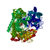 1unbC  1uobC  1uofC 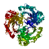 1uogC  1rxfS S: Starting model for refinement C: citing same article ( |
|---|---|
| Similar structure data |

-Links
 PDBj
PDBj


- Assembly
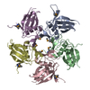
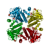
| Deposited unit | 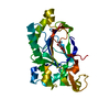
| ||||||||
|---|---|---|---|---|---|---|---|---|---|
| 1 | 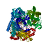
| ||||||||
| Unit cell |
|
-Components
| #1: Protein | Mass: 34591.641 Da / Num. of mol.: 1 Source method: isolated from a genetically manipulated source Source: (gene. exp.) STREPTOMYCES CLAVULIGERUS (bacteria) / Plasmid: PET11A / Production host: References: UniProt: P18548, deacetoxycephalosporin-C synthase |
|---|---|
| #2: Chemical | ChemComp-FE2 /   Mass: 55.845 Da / Num. of mol.: 1 / Source method: obtained synthetically / Formula: Fe Mass: 55.845 Da / Num. of mol.: 1 / Source method: obtained synthetically / Formula: Fe |
| #3: Chemical | ChemComp-SIN /   Mass: 118.088 Da / Num. of mol.: 1 / Source method: obtained synthetically / Formula: C4H6O4 Mass: 118.088 Da / Num. of mol.: 1 / Source method: obtained synthetically / Formula: C4H6O4 |
| #4: Water | ChemComp-HOH /  Mass: 18.015 Da / Num. of mol.: 216 / Source method: isolated from a natural source / Formula: H2O Mass: 18.015 Da / Num. of mol.: 216 / Source method: isolated from a natural source / Formula: H2O |
-Experimental details
-Experiment
| Experiment | Method: X-RAY DIFFRACTION / Number of used crystals: 1 |
|---|
- Sample preparation
| Crystal | Density Matthews: 2.6 Å3/Da / Density % sol: 53 % Description: THE DATA WERE COLLECTED FROM A MEROHEDRALLY TWINNED CRYSTAL | ||||||||||||||||||||
|---|---|---|---|---|---|---|---|---|---|---|---|---|---|---|---|---|---|---|---|---|---|
| Crystal grow | pH: 7.5 Details: 1.75M AMMONIUM SULPHATE, 5MM 2-OXOGLUTARATE,0.1M HEPES PH7.0, pH 7.50 | ||||||||||||||||||||
| Crystal grow | *PLUS Method: unknown / Details: Valegard, K., (1998) Nature, 394, 805. / PH range low: 7.5 / PH range high: 7 | ||||||||||||||||||||
| Components of the solutions | *PLUS
|
-Data collection
| Diffraction | Mean temperature: 100 K |
|---|---|
| Diffraction source | Source: SYNCHROTRON / Site: ESRF  / Beamline: BM14 / Wavelength: 0.8 / Beamline: BM14 / Wavelength: 0.8 |
| Detector | Type: MARRESEARCH / Detector: IMAGE PLATE / Date: Feb 15, 1998 |
| Radiation | Protocol: SINGLE WAVELENGTH / Monochromatic (M) / Laue (L): M / Scattering type: x-ray |
| Radiation wavelength | Wavelength: 0.8 Å / Relative weight: 1 |
| Reflection | Resolution: 1.5→56 Å / Num. obs: 45627 / % possible obs: 95.5 % / Redundancy: 2.5 % / Rmerge(I) obs: 0.049 |
| Reflection shell | Resolution: 1.5→1.55 Å / Redundancy: 2.2 % / Rmerge(I) obs: 0.28 / Mean I/σ(I) obs: 3.2 / % possible all: 94.2 |
| Reflection | *PLUS Highest resolution: 1.5 Å / Num. measured all: 108054 / Rmerge(I) obs: 0.049 |
| Reflection shell | *PLUS % possible obs: 94.2 % / Rmerge(I) obs: 0.28 |
- Processing
| Software |
| ||||||||||||||||||||
|---|---|---|---|---|---|---|---|---|---|---|---|---|---|---|---|---|---|---|---|---|---|
| Refinement | Method to determine structure: MOLECULAR REPLACEMENT Starting model: PDB ENTRY 1RXF Resolution: 1.5→55.9 Å / SU B: 2.008 / SU ML: 0.075 / Cross valid method: THROUGHOUT / ESU R: 0.078 / ESU R Free: 0.074 Details: REFINED AGAINST DETWINNED DATA THE RESIDUES MISSING IN THE PDB ENTRY (82-97,167-177 AND 310-311) ARE DISORDERED, AND THEREFORE OMITTED FROM THE MODEL
| ||||||||||||||||||||
| Displacement parameters | Biso mean: 19.8 Å2
| ||||||||||||||||||||
| Refinement step | Cycle: LAST / Resolution: 1.5→55.9 Å
| ||||||||||||||||||||
| Refinement | *PLUS Rfactor Rwork: 0.174 | ||||||||||||||||||||
| Solvent computation | *PLUS | ||||||||||||||||||||
| Displacement parameters | *PLUS |
