 Movie
Movie Controller

[English] 日本語
 Yorodumi
- PDB-1unr: Crystal structure of the PH domain of PKB alpha in complex with a... -
+ Open data

- Basic information
| Entry | Database: PDB / ID: 1unr | ||||||
|---|---|---|---|---|---|---|---|
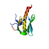
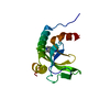
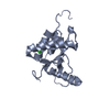
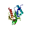
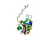
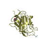
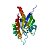
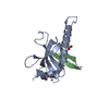
| Title | Crystal structure of the PH domain of PKB alpha in complex with a sulfate molecule | ||||||
 Components Components | RAC-ALPHA SERINE/THREONINE KINASE | ||||||
 Keywords Keywords | TRANSFERASE / PLECKSTRIN HOMOLOGY / PH / PKB / ATK / PHOSPHOINOSITIDE | ||||||
| Function / homology |  Function and homology information Function and homology informationpositive regulation of endodeoxyribonuclease activity / regulation of tRNA methylation / negative regulation of protein maturation / negative regulation of fatty acid beta-oxidation / positive regulation of protein localization to endoplasmic reticulum / regulation of glycogen biosynthetic process / negative regulation of lymphocyte migration / cellular response to decreased oxygen levels / cellular response to rapamycin / negative regulation of protein localization to lysosome ...positive regulation of endodeoxyribonuclease activity / regulation of tRNA methylation / negative regulation of protein maturation / negative regulation of fatty acid beta-oxidation / positive regulation of protein localization to endoplasmic reticulum / regulation of glycogen biosynthetic process / negative regulation of lymphocyte migration / cellular response to decreased oxygen levels / cellular response to rapamycin / negative regulation of protein localization to lysosome / maternal placenta development / maintenance of protein location in mitochondrion / mammalian oogenesis stage / AKT-mediated inactivation of FOXO1A / negative regulation of long-chain fatty acid import across plasma membrane / Negative regulation of the PI3K/AKT network / regulation of type B pancreatic cell development / positive regulation of anaphase-promoting complex-dependent catabolic process / : / potassium channel activator activity / sperm glycocalyx / AKT phosphorylates targets in the nucleus / activation-induced cell death of T cells / mammary gland epithelial cell differentiation / cellular response to oxidised low-density lipoprotein particle stimulus / negative regulation of cilium assembly / negative regulation of hydrogen peroxide-induced neuron intrinsic apoptotic signaling pathway / Butyrate Response Factor 1 (BRF1) binds and destabilizes mRNA / fibroblast migration / positive regulation of TORC2 signaling / positive regulation of glucose metabolic process / perinuclear theca / RUNX2 regulates genes involved in cell migration / cellular response to peptide / beta-arrestin-dependent dopamine receptor signaling pathway / positive regulation of organ growth / interleukin-18-mediated signaling pathway / positive regulation of sodium ion transport / MTOR signalling / response to growth factor / cell migration involved in sprouting angiogenesis / peripheral nervous system myelin maintenance / response to fluid shear stress / cellular response to granulocyte macrophage colony-stimulating factor stimulus / negative regulation of leukocyte cell-cell adhesion / RAB GEFs exchange GTP for GDP on RABs / glycogen biosynthetic process / phosphatidylinositol-3,4-bisphosphate binding / positive regulation of protein localization to cell surface / phosphorylation / complement receptor mediated signaling pathway / sphingosine-1-phosphate receptor signaling pathway / response to growth hormone / AKT phosphorylates targets in the cytosol / anoikis / regulation of postsynapse organization / positive regulation of fibroblast migration / labyrinthine layer blood vessel development / execution phase of apoptosis / regulation of myelination / KSRP (KHSRP) binds and destabilizes mRNA / response to UV-A / Regulation of TP53 Activity through Association with Co-factors / TORC2 complex binding / Mechanical load activates signaling by PIEZO1 and integrins in osteocytes / negative regulation of macroautophagy / Co-inhibition by CTLA4 / negative regulation of release of cytochrome c from mitochondria / negative regulation of cGAS/STING signaling pathway / peptidyl-threonine phosphorylation / cellular response to stress / response to food / regulation of neuron projection development / negative regulation of PERK-mediated unfolded protein response / Constitutive Signaling by AKT1 E17K in Cancer / apoptotic mitochondrial changes / positive regulation of protein metabolic process / phosphatidylinositol-3,4,5-trisphosphate binding / Regulation of localization of FOXO transcription factors / negative regulation of Notch signaling pathway / TOR signaling / cellular response to vascular endothelial growth factor stimulus / behavioral response to pain / CD28 dependent PI3K/Akt signaling / positive regulation of peptidyl-serine phosphorylation / Activation of BAD and translocation to mitochondria / positive regulation of blood vessel endothelial cell migration / Estrogen-dependent nuclear events downstream of ESR-membrane signaling / positive regulation of glycogen biosynthetic process / negative regulation of extrinsic apoptotic signaling pathway in absence of ligand / Mitochondrial unfolded protein response (UPRmt) / protein serine/threonine kinase inhibitor activity / response to insulin-like growth factor stimulus / SARS-CoV-2 targets host intracellular signalling and regulatory pathways / positive regulation of lipid biosynthetic process / positive regulation of fat cell differentiation / negative regulation of oxidative stress-induced intrinsic apoptotic signaling pathway / Cyclin E associated events during G1/S transition / positive regulation of G1/S transition of mitotic cell cycle / vascular endothelial cell response to laminar fluid shear stress Similarity search - Function | ||||||
| Biological species |  HOMO SAPIENS (human) HOMO SAPIENS (human) | ||||||
| Method |  X-RAY DIFFRACTION / SYNCHROTRON / MAD / Resolution: 1.25 Å X-RAY DIFFRACTION / SYNCHROTRON / MAD / Resolution: 1.25 Å | ||||||
 Authors Authors | Milburn, C.C. / Deak, M. / Kelly, S.M. / Price, N.C. / Alessi, D.R. / van Aalten, D.M.F. | ||||||
 Citation Citation | Journal: Biochem. J. / Year: 2003 Title: Binding of phosphatidylinositol 3,4,5-trisphosphate to the pleckstrin homology domain of protein kinase B induces a conformational change. Authors: Milburn, C.C. / Deak, M. / Kelly, S.M. / Price, N.C. / Alessi, D.R. / Van Aalten, D.M. #1: Journal: Curr.Biol. / Year: 2002Title: High Resolution Structure of the Pleckstrin Homology Domain of Protein Kinase B/Akt Bound to Phosphatidylinositol (3,4,5)-Trisphosphate Authors: Milburn, C.C. / Deak, M. / Alessi, D.R. / Van Aalten, D.M.F. | ||||||
| History |
|
- Structure visualization
| Structure viewer | Molecule: MolmilJmol/JSmol |
|---|
- Downloads & links
-Download
| PDBx/mmCIF format | 1unr.cif.gz | 66 KB | Display | PDBx/mmCIF format |
|---|---|---|---|---|
| PDB format | pdb1unr.ent.gz | 47.7 KB | Display | PDB format |
| PDBx/mmJSON format | 1unr.json.gz | Tree view | PDBx/mmJSON format | |
| Others |  Other downloads Other downloads |
-Validation report
| Arichive directory | https://data.pdbj.org/pub/pdb/validation_reports/un/1unrftp://data.pdbj.org/pub/pdb/validation_reports/un/1unr | HTTPS FTP |
|---|
-Related structure data
| Related structure data |  1unpC 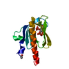 1unqC 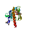 1h10S S: Starting model for refinement C: citing same article ( |
|---|---|
| Similar structure data |


-Links
 PDBj
PDBj


- Assembly
| Deposited unit | 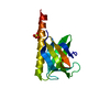
| ||||||||
|---|---|---|---|---|---|---|---|---|---|
| 1 |
| ||||||||
| Unit cell |
|
-Components
| #1: Protein | Mass: 14799.705 Da / Num. of mol.: 1 / Fragment: PLECKSTRIN HOMOLOGY DOMAIN, RESIDUES 1-123 Source method: isolated from a genetically manipulated source Source: (gene. exp.) HOMO SAPIENS (human) / Plasmid: PGEX4T-1 / Production host:  References: UniProt: P31749, non-specific serine/threonine protein kinase |
|---|---|
| #2: Chemical | ChemComp-SO4 /   Mass: 96.063 Da / Num. of mol.: 1 / Source method: obtained synthetically / Formula: SO4 Mass: 96.063 Da / Num. of mol.: 1 / Source method: obtained synthetically / Formula: SO4 |
| #3: Water | ChemComp-HOH /  Mass: 18.015 Da / Num. of mol.: 62 / Source method: isolated from a natural source / Formula: H2O Mass: 18.015 Da / Num. of mol.: 62 / Source method: isolated from a natural source / Formula: H2O |
| Has protein modification | Y |
-Experimental details
-Experiment
| Experiment | Method: X-RAY DIFFRACTION / Number of used crystals: 1 |
|---|
- Sample preparation
| Crystal | Density Matthews: 1.44 Å3/Da / Density % sol: 30 % |
|---|---|
| Crystal grow | pH: 8.5 / Details: 0.1 M TRIS (8.5) 0.2 M AMMONIUM SULFATE, pH 8.50 |
-Data collection
| Diffraction | Mean temperature: 100 K |
|---|---|
| Diffraction source | Source: SYNCHROTRON / Site: ESRF  / Beamline: ID14-2 / Wavelength: 0.933 / Beamline: ID14-2 / Wavelength: 0.933 |
| Detector | Date: Jan 15, 2002 |
| Radiation | Protocol: SINGLE WAVELENGTH / Monochromatic (M) / Laue (L): M / Scattering type: x-ray |
| Radiation wavelength | Wavelength: 0.933 Å / Relative weight: 1 |
| Reflection | Resolution: 1.25→15 Å / Num. obs: 27697 / % possible obs: 96.2 % / Redundancy: 4.3 % / Rmerge(I) obs: 0.073 / Net I/σ(I): 21.5 |
| Reflection shell | Resolution: 1.25→1.29 Å / Redundancy: 2.4 % / Rmerge(I) obs: 0.394 / Mean I/σ(I) obs: 2.7 / % possible all: 74.6 |
- Processing
| Software |
| |||||||||||||||||||||||||||||||||
|---|---|---|---|---|---|---|---|---|---|---|---|---|---|---|---|---|---|---|---|---|---|---|---|---|---|---|---|---|---|---|---|---|---|---|
| Refinement | Method to determine structure: MAD Starting model: PDB ENTRY 1H10 Resolution: 1.25→20 Å / Num. parameters: 9408 / Num. restraintsaints: 11985 / Cross valid method: THROUGHOUT / σ(F): 0 / Stereochemistry target values: ENGH AND HUBER
| |||||||||||||||||||||||||||||||||
| Solvent computation | Solvent model: MOEWS & KRETSINGER, J.MOL.BIOL.91(1973)201-2 | |||||||||||||||||||||||||||||||||
| Refine analyze | Num. disordered residues: 6 / Occupancy sum hydrogen: 767.7 / Occupancy sum non hydrogen: 995.24 | |||||||||||||||||||||||||||||||||
| Refinement step | Cycle: LAST / Resolution: 1.25→20 Å
| |||||||||||||||||||||||||||||||||
| Refine LS restraints |
|
