[English] 日本語
 Yorodumi
- PDB-1une: CARBOXYLIC ESTER HYDROLASE, 1.5 ANGSTROM ORTHORHOMBIC FORM OF THE... -
+ Open data
- Basic information
| Entry | Database: PDB / ID: 1une | ||||||
|---|---|---|---|---|---|---|---|
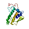
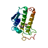
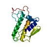
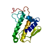
| Title | CARBOXYLIC ESTER HYDROLASE, 1.5 ANGSTROM ORTHORHOMBIC FORM OF THE BOVINE RECOMBINANT PLA2 | ||||||
 Components Components | PHOSPHOLIPASE A2 | ||||||
 Keywords Keywords | HYDROLASE / ENZYME / CARBOXYLIC ESTER HYDROLASE | ||||||
| Function / homology |  Function and homology information Function and homology informationAcyl chain remodelling of PS / Acyl chain remodelling of PG / Synthesis of PA / Acyl chain remodelling of PC / Acyl chain remodelling of PE / Acyl chain remodelling of PI / positive regulation of podocyte apoptotic process / phosphatidylglycerol metabolic process / phosphatidylcholine metabolic process / phospholipase A2 ...Acyl chain remodelling of PS / Acyl chain remodelling of PG / Synthesis of PA / Acyl chain remodelling of PC / Acyl chain remodelling of PE / Acyl chain remodelling of PI / positive regulation of podocyte apoptotic process / phosphatidylglycerol metabolic process / phosphatidylcholine metabolic process / phospholipase A2 / bile acid binding / calcium-dependent phospholipase A2 activity / arachidonate secretion / lipid catabolic process / innate immune response in mucosa / phospholipid binding / antimicrobial humoral immune response mediated by antimicrobial peptide / positive regulation of fibroblast proliferation / fatty acid biosynthetic process / antibacterial humoral response / defense response to Gram-positive bacterium / signaling receptor binding / calcium ion binding / cell surface / extracellular space Similarity search - Function | ||||||
| Biological species |  | ||||||
| Method |  X-RAY DIFFRACTION / THE HIGH RESOLUTION ATOMIC COORDINATES OF THE WILD TYPE (PDB ENTRY 1BP2) WERE USED AS THE STARTING MODEL FOR REFINEMENT. / Resolution: 1.5 Å X-RAY DIFFRACTION / THE HIGH RESOLUTION ATOMIC COORDINATES OF THE WILD TYPE (PDB ENTRY 1BP2) WERE USED AS THE STARTING MODEL FOR REFINEMENT. / Resolution: 1.5 Å | ||||||
 Authors Authors | Sundaralingam, M. | ||||||
 Citation Citation |  Journal: Acta Crystallogr.,Sect.D / Year: 1999 Journal: Acta Crystallogr.,Sect.D / Year: 1999Title: High-resolution refinement of orthorhombic bovine pancreatic phospholipase A2. Authors: Sekar, K. / Sundaralingam, M. #1:  Journal: To be Published Journal: To be PublishedTitle: Crystal Structure of the Complex of Bovine Pancreatic Phospholipase A2 with a Transition State Analogue Authors: Sekar, K. / Kumar, A. / Liu, X. / Tsai, M.-D. / Gelb, M.H. / Sundaralingam, M. #2:  Journal: To be Published Journal: To be PublishedTitle: 1.72A Resolution Refinement of the Trigonal Form of Bovine Pancreatic Phospholipase A2 Authors: Sekar, K. / Sekarudu, C. / Tsai, M.-D. / Sundaralingam, M. #3:  Journal: Biochemistry / Year: 1997 Journal: Biochemistry / Year: 1997Title: Crystal Structure of the Complex of Bovine Pancreatic Phospholipase A2 with the Inhibitor 1-Hexadecyl-3-(Trifluoroethyl)-Sn-Glycero-2-Phosphomethanol Authors: Sekar, K. / Eswaramoorthy, S. / Jain, M.K. / Sundaralingam, M. #4:  Journal: Biochemistry / Year: 1997 Journal: Biochemistry / Year: 1997Title: Phospholipase A2 Engineering. Structural and Functional Roles of the Highly Conserved Active Site Residue Aspartate-99 Authors: Sekar, K. / Yu, B.Z. / Rogers, J. / Lutton, J. / Liu, X. / Chen, X. / Tsai, M.D. / Jain, M.K. / Sundaralingam, M. #5:  Journal: Biochemistry / Year: 1996 Journal: Biochemistry / Year: 1996Title: Phospholipase A2 Engineering. Deletion of the C-Terminus Segment Changes Substrate Specificity and Uncouples Calcium and Substrate Binding at the Zwitterionic Interface Authors: Huang, B. / Yu, B.Z. / Rogers, J. / Byeon, I.J. / Sekar, K. / Chen, X. / Sundaralingam, M. / Tsai, M.D. / Jain, M.K. #6:  Journal: Biochemistry / Year: 1991 Journal: Biochemistry / Year: 1991Title: Phospholipase A2 Engineering. X-Ray Structural and Functional Evidence for the Interaction of Lysine-56 with Substrates Authors: Noel, J.P. / Bingman, C.A. / Deng, T.L. / Dupureur, C.M. / Hamilton, K.J. / Jiang, R.T. / Kwak, J.G. / Sekharudu, C. / Sundaralingam, M. / Tsai, M.D. | ||||||
| History |
|
- Structure visualization
| Structure viewer | Molecule:  Molmil Molmil Jmol/JSmol Jmol/JSmol |
|---|
- Downloads & links
- Download
| PDBx/mmCIF format |  1une.cif.gz 1une.cif.gz | 40.6 KB | Display |  PDBx/mmCIF format PDBx/mmCIF format |
|---|---|---|---|---|
| PDB format |  pdb1une.ent.gz pdb1une.ent.gz | 27.1 KB | Display |  PDB format PDB format |
| PDBx/mmJSON format |  1une.json.gz 1une.json.gz | Tree view |  PDBx/mmJSON format PDBx/mmJSON format | |
| Others |  Other downloads Other downloads |
-Validation report
| Arichive directory |  https://data.pdbj.org/pub/pdb/validation_reports/un/1une https://data.pdbj.org/pub/pdb/validation_reports/un/1une ftp://data.pdbj.org/pub/pdb/validation_reports/un/1une ftp://data.pdbj.org/pub/pdb/validation_reports/un/1une | HTTPS FTP |
|---|
-Related structure data
| Related structure data |  1bp2S S: Starting model for refinement |
|---|---|
| Similar structure data |
- Links
Links
- Assembly
| Deposited unit | 
| ||||||||
|---|---|---|---|---|---|---|---|---|---|
| 1 |
| ||||||||
| Unit cell |
|
- Components
| #1: Protein | Mass: 13810.504 Da / Num. of mol.: 1 Source method: isolated from a genetically manipulated source Source: (gene. exp.)   |
|---|---|
| #2: Chemical | ChemComp-CA / |
| #3: Water | ChemComp-HOH / |
| Has protein modification | Y |
-Experimental details
-Experiment
| Experiment | Method:  X-RAY DIFFRACTION / Number of used crystals: 1 X-RAY DIFFRACTION / Number of used crystals: 1 |
|---|
- Sample preparation
| Crystal | Density Matthews: 2.1 Å3/Da / Density % sol: 41.44 % | ||||||||||||||||||||||||||||||||||||
|---|---|---|---|---|---|---|---|---|---|---|---|---|---|---|---|---|---|---|---|---|---|---|---|---|---|---|---|---|---|---|---|---|---|---|---|---|---|
| Crystal grow | Method: vapor diffusion, hanging drop / pH: 7.2 Details: CRYSTALS WERE GROWN BY CO-CRYSTALLIZATION BY THE HANGING DROP VAPOR DIFFUSION METHOD USING THE CONDITIONS 5 (MICRO)L OF THE PROTEIN (15 MG/ML OF THE PROTEIN), 5MM CACL2, 50MM TRIS BUFFER, PH ...Details: CRYSTALS WERE GROWN BY CO-CRYSTALLIZATION BY THE HANGING DROP VAPOR DIFFUSION METHOD USING THE CONDITIONS 5 (MICRO)L OF THE PROTEIN (15 MG/ML OF THE PROTEIN), 5MM CACL2, 50MM TRIS BUFFER, PH 7.2 AND 3.0 (MICRO)L OF 50% MPD IN THE DROPLET. THE RESERVOIR CONTAINED (50%) OF MPD., vapor diffusion - hanging drop | ||||||||||||||||||||||||||||||||||||
| Crystal grow | *PLUS Method: vapor diffusion, hanging drop | ||||||||||||||||||||||||||||||||||||
| Components of the solutions | *PLUS
|
-Data collection
| Diffraction | Mean temperature: 291 K |
|---|---|
| Diffraction source | Source:  ROTATING ANODE / Type: RIGAKU R-AXIS II / Wavelength: 1.5418 ROTATING ANODE / Type: RIGAKU R-AXIS II / Wavelength: 1.5418 |
| Detector | Type: RIGAKU RAXIS IIC / Detector: IMAGE PLATE / Date: Jan 26, 1996 |
| Radiation | Monochromator: GRAPHITE(002) / Monochromatic (M) / Laue (L): M / Scattering type: x-ray |
| Radiation wavelength | Wavelength: 1.5418 Å / Relative weight: 1 |
| Reflection | Resolution: 1.5→10 Å / Num. obs: 17572 / % possible obs: 92 % / Redundancy: 3 % / Rmerge(I) obs: 0.046 |
| Reflection shell | Resolution: 1.5→1.55 Å / Redundancy: 3.7 % / Rmerge(I) obs: 0.172 / % possible all: 63 |
| Reflection | *PLUS Num. measured all: 68591 |
| Reflection shell | *PLUS Num. unique obs: 1176 |
- Processing
| Software |
| ||||||||||||||||||||||||||||||||||||||||||||||||||||||||||||
|---|---|---|---|---|---|---|---|---|---|---|---|---|---|---|---|---|---|---|---|---|---|---|---|---|---|---|---|---|---|---|---|---|---|---|---|---|---|---|---|---|---|---|---|---|---|---|---|---|---|---|---|---|---|---|---|---|---|---|---|---|---|
| Refinement | Method to determine structure: THE HIGH RESOLUTION ATOMIC COORDINATES OF THE WILD TYPE (PDB ENTRY 1BP2) WERE USED AS THE STARTING MODEL FOR REFINEMENT. Starting model: WILD TYPE (PDB ENTRY 1BP2) Resolution: 1.5→10 Å / Rfactor Rfree error: 0.24 / Data cutoff high absF: 0.1 / Data cutoff low absF: 1000000 / σ(F): 1
| ||||||||||||||||||||||||||||||||||||||||||||||||||||||||||||
| Refine analyze | Luzzati coordinate error obs: 0.19 Å / Luzzati sigma a obs: 0.21 Å | ||||||||||||||||||||||||||||||||||||||||||||||||||||||||||||
| Refinement step | Cycle: LAST / Resolution: 1.5→10 Å
| ||||||||||||||||||||||||||||||||||||||||||||||||||||||||||||
| Refine LS restraints |
| ||||||||||||||||||||||||||||||||||||||||||||||||||||||||||||
| LS refinement shell | Resolution: 1.5→1.55 Å / Total num. of bins used: 8
| ||||||||||||||||||||||||||||||||||||||||||||||||||||||||||||
| Xplor file |
| ||||||||||||||||||||||||||||||||||||||||||||||||||||||||||||
| Software | *PLUS Name:  X-PLOR / Version: 3.1 / Classification: refinement X-PLOR / Version: 3.1 / Classification: refinement | ||||||||||||||||||||||||||||||||||||||||||||||||||||||||||||
| Refine LS restraints | *PLUS
| ||||||||||||||||||||||||||||||||||||||||||||||||||||||||||||
| LS refinement shell | *PLUS Rfactor Rwork: 0.34 |
 Movie
Movie Controller


 PDBj
PDBj


