 Movie
Movie Controller

[English] 日本語
 Yorodumi
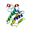
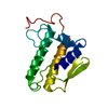
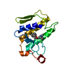
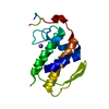
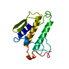
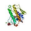
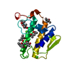
- PDB-1bp2: STRUCTURE OF BOVINE PANCREATIC PHOSPHOLIPASE A2 AT 1.7 ANGSTROMS ... -
+ Open data

- Basic information
| Entry | Database: PDB / ID: 1bp2 | ||||||
|---|---|---|---|---|---|---|---|
| Title | STRUCTURE OF BOVINE PANCREATIC PHOSPHOLIPASE A2 AT 1.7 ANGSTROMS RESOLUTION | ||||||
 Components Components | PHOSPHOLIPASE A2 | ||||||
 Keywords Keywords | HYDROLASE | ||||||
| Function / homology |  Function and homology information Function and homology informationAcyl chain remodelling of PS / Acyl chain remodelling of PG / Synthesis of PA / Acyl chain remodelling of PC / Acyl chain remodelling of PE / Acyl chain remodelling of PI / positive regulation of podocyte apoptotic process / phosphatidylglycerol metabolic process / phosphatidylcholine metabolic process / : ...Acyl chain remodelling of PS / Acyl chain remodelling of PG / Synthesis of PA / Acyl chain remodelling of PC / Acyl chain remodelling of PE / Acyl chain remodelling of PI / positive regulation of podocyte apoptotic process / phosphatidylglycerol metabolic process / phosphatidylcholine metabolic process / : / bile acid binding / phospholipase A2 / arachidonate secretion / lipid catabolic process / innate immune response in mucosa / phospholipid binding / positive regulation of fibroblast proliferation / fatty acid biosynthetic process / antimicrobial humoral immune response mediated by antimicrobial peptide / antibacterial humoral response / defense response to Gram-positive bacterium / signaling receptor binding / calcium ion binding / cell surface / : Similarity search - Function | ||||||
| Biological species |  | ||||||
| Method |  X-RAY DIFFRACTION / Resolution: 1.7 Å X-RAY DIFFRACTION / Resolution: 1.7 Å | ||||||
 Authors Authors | Dijkstra, B.W. / Kalk, K.H. / Hol, W.G.J. / Drenth, J. | ||||||
 Citation Citation | Journal: J.Mol.Biol. / Year: 1981 Title: Structure of bovine pancreatic phospholipase A2 at 1.7A resolution. Authors: Dijkstra, B.W. / Kalk, K.H. / Hol, W.G. / Drenth, J. #1: Journal: J.Biol.Chem. / Year: 1985Title: A Comparison of the Crystal Structures of Phospholipase A2 from Bovine Pancreas and Crotalus Atrox Venom Authors: Renetseder, R. / Brunie, S. / Dijkstra, B.W. / Drenth, J. / Sigler, P.B. #2: Journal: Nature / Year: 1981Title: Active Site and Catalytic Mechanism of Phospholipase A2 Authors: Dijkstra, B.W. / Drenth, J. / Kalk, K.H. #3: Journal: Thesis / Year: 1980Title: Structure and Mechanism of Phospholipase A2 Authors: Dijkstra, B.W. #4: Journal: J.Mol.Biol. / Year: 1978Title: Three-Dimensional Structure and Disulfide Bond Connections in Bovine Pancreatic Phospholipase A2 Authors: Dijkstra, B.W. / Drenth, J. / Kalk, K.H. / Vandermaelen, P. | ||||||
| History |
|
- Structure visualization
| Structure viewer | Molecule: MolmilJmol/JSmol |
|---|
- Downloads & links
-Download
| PDBx/mmCIF format | 1bp2.cif.gz | 39.9 KB | Display | PDBx/mmCIF format |
|---|---|---|---|---|
| PDB format | pdb1bp2.ent.gz | 27 KB | Display | PDB format |
| PDBx/mmJSON format | 1bp2.json.gz | Tree view | PDBx/mmJSON format | |
| Others |  Other downloads Other downloads |
-Validation report
| Arichive directory | https://data.pdbj.org/pub/pdb/validation_reports/bp/1bp2ftp://data.pdbj.org/pub/pdb/validation_reports/bp/1bp2 | HTTPS FTP |
|---|
-Related structure data
| Similar structure data |
|---|


-Links
 PDBj
PDBj

- Assembly
| Deposited unit | 
| ||||||||
|---|---|---|---|---|---|---|---|---|---|
| 1 |
| ||||||||
| Unit cell |
|
-Components
| #1: Protein | Mass: 13810.504 Da / Num. of mol.: 1 Source method: isolated from a genetically manipulated source Source: (gene. exp.) | ||||||
|---|---|---|---|---|---|---|---|
| #2: Chemical | ChemComp-CA /   Mass: 40.078 Da / Num. of mol.: 1 / Source method: obtained synthetically / Formula: Ca Mass: 40.078 Da / Num. of mol.: 1 / Source method: obtained synthetically / Formula: Ca | ||||||
| #3: Chemical |   Mass: 118.174 Da / Num. of mol.: 2 / Source method: obtained synthetically / Formula: C6H14O2 / Comment: precipitant*YM Mass: 118.174 Da / Num. of mol.: 2 / Source method: obtained synthetically / Formula: C6H14O2 / Comment: precipitant*YM#4: Water | ChemComp-HOH / |  Mass: 18.015 Da / Num. of mol.: 106 / Source method: isolated from a natural source / Formula: H2O Mass: 18.015 Da / Num. of mol.: 106 / Source method: isolated from a natural source / Formula: H2OCompound details | THE TYPE CLASSIFICATIONS GIVEN IN THE REMARK FIELD OF THE TURN RECORDS BELOW FOLLOWS THE ...THE TYPE CLASSIFICA | Has protein modification | Y | |
-Experimental details
-Experiment
| Experiment | Method: X-RAY DIFFRACTION |
|---|
- Sample preparation
| Crystal | Density Matthews: 2.09 Å3/Da / Density % sol: 41.27 % |
|---|---|
| Crystal | *PLUS Density % sol: 39 % |
| Crystal grow | *PLUS Method: unknown |
-Data collection
| Radiation | Scattering type: x-ray |
|---|---|
| Radiation wavelength | Relative weight: 1 |
| Reflection | *PLUS Highest resolution: 1.7 Å / Num. all: 13534 / Num. obs: 11596 / % possible obs: 85.7 % / Observed criterion σ(I): 2 / Rmerge(I) obs: 0.025 |
- Processing
| Software | Name: AGARWAL / Version: FAST-FOURIER TRANSFORM LEAST-SQUARES PROCEDURE / Classification: refinement | ||||||||||||||||||||||||||||||||||||||||||||||||||||||||||||
|---|---|---|---|---|---|---|---|---|---|---|---|---|---|---|---|---|---|---|---|---|---|---|---|---|---|---|---|---|---|---|---|---|---|---|---|---|---|---|---|---|---|---|---|---|---|---|---|---|---|---|---|---|---|---|---|---|---|---|---|---|---|
| Refinement | Rfactor Rwork: 0.171 / Highest resolution: 1.7 Å | ||||||||||||||||||||||||||||||||||||||||||||||||||||||||||||
| Refinement step | Cycle: LAST / Highest resolution: 1.7 Å
| ||||||||||||||||||||||||||||||||||||||||||||||||||||||||||||
| Refine LS restraints |
| ||||||||||||||||||||||||||||||||||||||||||||||||||||||||||||
| Refinement | *PLUS Lowest resolution: 10 Å / Rfactor obs: 0.171 | ||||||||||||||||||||||||||||||||||||||||||||||||||||||||||||
| Solvent computation | *PLUS | ||||||||||||||||||||||||||||||||||||||||||||||||||||||||||||
| Displacement parameters | *PLUS | ||||||||||||||||||||||||||||||||||||||||||||||||||||||||||||
| Refine LS restraints | *PLUS Type: o_bond_d / Dev ideal: 0.005 |
