- PDB-2zp3: Carboxylic ester hydrolase, single mutant d49n of bovine pancreat... -
+
Open data
ID or keywords:
Loading...
-
Basic information
Entry
Database: PDB / ID: 2zp3
Title
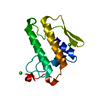
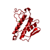
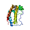
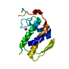
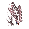
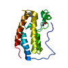
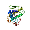
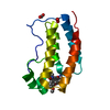
Carboxylic ester hydrolase, single mutant d49n of bovine pancreatic pla2 enzyme
Components
Phospholipase A2
Keywords
HYDROLASE / ACTIVE SITE MUTANT / METAL BINDING PROTEIN / Calcium / Lipid degradation / Metal-binding / Pyrrolidone carboxylic acid / Secreted
Function / homology
Function and homology information
Acyl chain remodelling of PS / Acyl chain remodelling of PG / Synthesis of PA / Acyl chain remodelling of PC / Acyl chain remodelling of PE / Acyl chain remodelling of PI / positive regulation of podocyte apoptotic process / phosphatidylglycerol metabolic process / phosphatidylcholine metabolic process / : ...Acyl chain remodelling of PS / Acyl chain remodelling of PG / Synthesis of PA / Acyl chain remodelling of PC / Acyl chain remodelling of PE / Acyl chain remodelling of PI / positive regulation of podocyte apoptotic process / phosphatidylglycerol metabolic process / phosphatidylcholine metabolic process / : / bile acid binding / phospholipase A2 / arachidonate secretion / lipid catabolic process / innate immune response in mucosa / phospholipid binding / positive regulation of fibroblast proliferation / fatty acid biosynthetic process / antimicrobial humoral immune response mediated by antimicrobial peptide / antibacterial humoral response / defense response to Gram-positive bacterium / signaling receptor binding / calcium ion binding / cell surface / : Similarity search - Function
Phospholipase A2, aspartic acid active site / Phospholipase A2 aspartic acid active site. / Phospholipase A2, histidine active site / Phospholipase A2 histidine active site. / Phospholipase A2 / Phospholipase A2 domain / Phospholipase A2 / Phospholipase A2 / Phospholipase A2 domain / Phospholipase A2 ...Phospholipase A2, aspartic acid active site / Phospholipase A2 aspartic acid active site. / Phospholipase A2, histidine active site / Phospholipase A2 histidine active site. / Phospholipase A2 / Phospholipase A2 domain / Phospholipase A2 / Phospholipase A2 / Phospholipase A2 domain / Phospholipase A2 / Phospholipase A2 domain superfamily / Up-down Bundle / Mainly Alpha Similarity search - Domain/homology
In the structure databanks used in Yorodumi, some data are registered as the other names, "COVID-19 virus" and "2019-nCoV". Here are the details of the virus and the list of structure data.
Jan 31, 2019. EMDB accession codes are about to change! (news from PDBe EMDB page)
EMDB accession codes are about to change! (news from PDBe EMDB page)
The allocation of 4 digits for EMDB accession codes will soon come to an end. Whilst these codes will remain in use, new EMDB accession codes will include an additional digit and will expand incrementally as the available range of codes is exhausted. The current 4-digit format prefixed with “EMD-” (i.e. EMD-XXXX) will advance to a 5-digit format (i.e. EMD-XXXXX), and so on. It is currently estimated that the 4-digit codes will be depleted around Spring 2019, at which point the 5-digit format will come into force.
The EM Navigator/Yorodumi systems omit the EMD- prefix.
Related info.:Q: What is EMD? / ID/Accession-code notation in Yorodumi/EM Navigator
Yorodumi is a browser for structure data from EMDB, PDB, SASBDB, etc.
This page is also the successor to EM Navigator detail page, and also detail information page/front-end page for Omokage search.
The word "yorodu" (or yorozu) is an old Japanese word meaning "ten thousand". "mi" (miru) is to see.
Related info.:EMDB / PDB / SASBDB / Comparison of 3 databanks / Yorodumi Search / Aug 31, 2016. New EM Navigator & Yorodumi / Yorodumi Papers / Jmol/JSmol / Function and homology information / Changes in new EM Navigator and Yorodumi
 Movie
Movie Controller

 Yorodumi
 Open data
 Basic information
 Components
 Keywords
 Function and homology information
 X-RAY DIFFRACTION /
  Authors
 Citation
 Structure visualization
 Downloads & links
 Other downloads
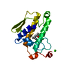


 PDBj
PDBj

 Assembly
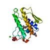


 Mass: 40.078 Da / Num. of mol.: 1 / Source method: obtained synthetically / Formula: Ca
 Mass: 35.453 Da / Num. of mol.: 1 / Source method: obtained synthetically / Formula: Cl
 Mass: 122.143 Da / Num. of mol.: 1 / Source method: obtained synthetically / Formula: C4H12NO3 / Comment: pH buffer*YM
 Mass: 118.174 Da / Num. of mol.: 1 / Source method: obtained synthetically / Formula: C6H14O2 / Comment: precipitant*YM
 Sample preparation
 Processing