 Movie
Movie Controller

[English] 日本語
 Yorodumi
- PDB-1um2: Crystal Structure of the Vma1-Derived Endonuclease with the Ligat... -
+ Open data

- Basic information
| Entry | Database: PDB / ID: 1um2 | ||||||
|---|---|---|---|---|---|---|---|
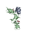
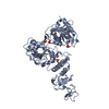
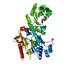
| Title | Crystal Structure of the Vma1-Derived Endonuclease with the Ligated Extein Segment | ||||||
 Components Components |
| ||||||
 Keywords Keywords | HYDROLASE / Protein splicing / Vma1-derived endonuclease / VDE / intein / extein / thiazolidine | ||||||
| Function / homology |  Function and homology information Function and homology informationInsulin receptor recycling / Transferrin endocytosis and recycling / ROS and RNS production in phagocytes / Golgi lumen acidification / Amino acids regulate mTORC1 / vacuolar proton-transporting V-type ATPase, V1 domain / endosomal lumen acidification / proton-transporting V-type ATPase complex / intron homing / vacuolar proton-transporting V-type ATPase complex ...Insulin receptor recycling / Transferrin endocytosis and recycling / ROS and RNS production in phagocytes / Golgi lumen acidification / Amino acids regulate mTORC1 / vacuolar proton-transporting V-type ATPase, V1 domain / endosomal lumen acidification / proton-transporting V-type ATPase complex / intron homing / vacuolar proton-transporting V-type ATPase complex / intein-mediated protein splicing / vacuolar acidification / fungal-type vacuole membrane / proton-transporting ATPase activity, rotational mechanism / H+-transporting two-sector ATPase / ATP metabolic process / proton transmembrane transport / endonuclease activity / Hydrolases; Acting on ester bonds / Golgi membrane / hydrolase activity / mRNA binding / DNA binding / ATP binding Similarity search - Function | ||||||
| Biological species |  | ||||||
| Method |  X-RAY DIFFRACTION / SYNCHROTRON / MOLECULAR REPLACEMENT / Resolution: 2.9 Å X-RAY DIFFRACTION / SYNCHROTRON / MOLECULAR REPLACEMENT / Resolution: 2.9 Å | ||||||
 Authors Authors | Mizutani, R. / Anraku, Y. / Satow, Y. | ||||||
 Citation Citation | Journal: J.Synchrotron Radiat. / Year: 2004 Title: Protein splicing of yeast VMA1-derived endonuclease via thiazolidine intermediates. Authors: Mizutani, R. / Anraku, Y. / Satow, Y. #1: Journal: J.Mol.Biol. / Year: 2002Title: Protein-splicing reaction via a thiazolidine intermediate: crystal structure of the VMA1-derived endonuclease bearing the N and C-terminal propeptides Authors: Mizutani, R. / Nogami, S. / Kawasaki, M. / Ohya, Y. / Anraku, Y. / Satow, Y. | ||||||
| History |
|
- Structure visualization
| Structure viewer | Molecule: MolmilJmol/JSmol |
|---|
- Downloads & links
-Download
| PDBx/mmCIF format | 1um2.cif.gz | 178.8 KB | Display | PDBx/mmCIF format |
|---|---|---|---|---|
| PDB format | pdb1um2.ent.gz | 142.1 KB | Display | PDB format |
| PDBx/mmJSON format | 1um2.json.gz | Tree view | PDBx/mmJSON format | |
| Others |  Other downloads Other downloads |
-Validation report
| Arichive directory | https://data.pdbj.org/pub/pdb/validation_reports/um/1um2ftp://data.pdbj.org/pub/pdb/validation_reports/um/1um2 | HTTPS FTP |
|---|
-Related structure data
| Related structure data | 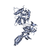 1jvaS S: Starting model for refinement |
|---|---|
| Similar structure data |


-Links
 PDBj
PDBj


- Assembly
| Deposited unit | 
| ||||||||
|---|---|---|---|---|---|---|---|---|---|
| 1 | 
| ||||||||
| 2 | 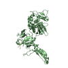
| ||||||||
| Unit cell |
|
-Components
| #1: Protein | Mass: 50963.789 Da / Num. of mol.: 2 / Mutation: C284S, H362N Source method: isolated from a genetically manipulated source Source: (gene. exp.) Gene: vma1 / Plasmid: pET-17b-VDE-X10SNS / Species (production host): Escherichia coli / Production host:  References: UniProt: P17255, H+-transporting two-sector ATPase #2: Protein/peptide | Mass: 2231.400 Da / Num. of mol.: 2 / Mutation: C738S Source method: isolated from a genetically manipulated source Details: The N-extein segments: residues 273-283 and C-extein segments: 738-747 were ligated by protein splicing. residue 283 links with residue 738 Source: (gene. exp.) Gene: vma1 / Plasmid: pET-17b-VDE-X10SNS / Species (production host): Escherichia coli / Production host: References: UniProt: P17255, H+-transporting two-sector ATPase #3: Water | ChemComp-HOH / |  Mass: 18.015 Da / Num. of mol.: 153 / Source method: isolated from a natural source / Formula: H2O Mass: 18.015 Da / Num. of mol.: 153 / Source method: isolated from a natural source / Formula: H2O |
|---|
-Experimental details
-Experiment
| Experiment | Method: X-RAY DIFFRACTION / Number of used crystals: 1 |
|---|
- Sample preparation
| Crystal | Density Matthews: 2.44 Å3/Da / Density % sol: 49.62 % |
|---|---|
| Crystal grow | Temperature: 293 K / Method: vapor diffusion, hanging drop / pH: 6.2 Details: PEG 6000, BistrisHCl, 2-mercaptoethanol, magnesium chloride, cadmium chloride, pH 6.2, VAPOR DIFFUSION, HANGING DROP, temperature 293K |
-Data collection
| Diffraction | Mean temperature: 110 K |
|---|---|
| Diffraction source | Source: SYNCHROTRON / Site: SPring-8  / Beamline: BL40B2 / Wavelength: 0.7 Å / Beamline: BL40B2 / Wavelength: 0.7 Å |
| Detector | Type: RIGAKU RAXIS IV++ / Detector: IMAGE PLATE / Date: Mar 17, 2000 Details: DOUBLE CRYSTAL MONOCHROMATOR AND BENT-CYLINDER MIRROR |
| Radiation | Monochromator: SI 111 / Protocol: SINGLE WAVELENGTH / Monochromatic (M) / Laue (L): M / Scattering type: x-ray |
| Radiation wavelength | Wavelength: 0.7 Å / Relative weight: 1 |
| Reflection | Resolution: 2.9→30 Å / Num. all: 20700 / Num. obs: 20700 / % possible obs: 83 % / Observed criterion σ(F): 0 / Observed criterion σ(I): 0 / Redundancy: 2.4 % / Rmerge(I) obs: 0.051 |
| Reflection shell | Resolution: 2.9→3 Å / Rmerge(I) obs: 0.141 / Num. unique all: 1519 / % possible all: 60.1 |
- Processing
| Software |
| ||||||||||||||||||||
|---|---|---|---|---|---|---|---|---|---|---|---|---|---|---|---|---|---|---|---|---|---|
| Refinement | Method to determine structure: MOLECULAR REPLACEMENT Starting model: PDB ENTRY 1jva Resolution: 2.9→30 Å / Isotropic thermal model: isotropic / Cross valid method: THROUGHOUT / σ(F): 0 / Stereochemistry target values: Engh & Huber
| ||||||||||||||||||||
| Refine analyze | Luzzati coordinate error obs: 0.34 Å / Luzzati d res low obs: 5 Å | ||||||||||||||||||||
| Refinement step | Cycle: LAST / Resolution: 2.9→30 Å
| ||||||||||||||||||||
| Refine LS restraints |
|
