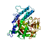
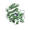
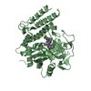
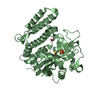
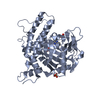
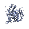
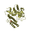
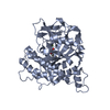
Entry Database : PDB / ID : 1uk1Title Crystal structure of human poly(ADP-ribose) polymerase complexed with a potent inhibitor Poly [ADP-ribose] polymerase-1 Keywords / Function / homology Function Domain/homology Component
/ / / / / / / / / / / / / / / / / / / / / / / / / / / / / / / / / / / / / / / / / / / / / / / / / / / / / / / / / / / / / / / / / / / / / / / / / / / / / / / / / / / / / / / / / / / / / / / / / / / / / / / / / / / / / / / / / / / / / / / / / / / / / / / / / / / / / / / / / / / / / / / / / / / / / / / / / / / / Biological species Homo sapiens (human)Method / / / Resolution : 3 Å Authors Kinoshita, T. Journal : J.Med.Chem. / Year : 2004Title : Rational approaches to discovery of orally active and brain-penetrable quinazolinone inhibitors of poly(ADP-ribose)polymeraseAuthors : Hattori, K. / Kido, Y. / Yamamoto, H. / Ishida, J. / Kamijo, K. / Murano, K. / Ohkubo, M. / Kinoshita, T. / Iwashita, A. / Mihara, K. / Yamazaki, S. / Matsuoka, N. / Teramura, Y. / Miyake, H. History Deposition Aug 14, 2003 Deposition site / Processing site Revision 1.0 Sep 14, 2004 Provider / Type Revision 1.1 Apr 27, 2008 Group Revision 1.2 Jul 13, 2011 Group Revision 1.3 Dec 27, 2023 Group / Database references / Derived calculationsCategory chem_comp_atom / chem_comp_bond ... chem_comp_atom / chem_comp_bond / database_2 / struct_site Item _database_2.pdbx_DOI / _database_2.pdbx_database_accession ... _database_2.pdbx_DOI / _database_2.pdbx_database_accession / _struct_site.pdbx_auth_asym_id / _struct_site.pdbx_auth_comp_id / _struct_site.pdbx_auth_seq_id
Show all Show less
 Movie
Movie Controller

 Yorodumi
 Open data
 Basic information
 Components
 Keywords
 Function and homology information
 Homo sapiens (human)
 X-RAY DIFFRACTION /
  Authors
 Citation
 Structure visualization
 Downloads & links
 Other downloads
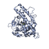


 PDBj
PDBj


 Assembly


 Mass: 393.454 Da / Num. of mol.: 2 / Source method: obtained synthetically / Formula: C23H24FN3O2
 Mass: 18.015 Da / Num. of mol.: 191 / Source method: isolated from a natural source / Formula: H2O
 Sample preparation
 / Beamline: BL-6B / Wavelength: 1 Å
 Processing