 Movie
Movie Controller

[English] 日本語
 Yorodumi
- PDB-1ujn: Crystal structure of dehydroquinate synthase from Thermus thermop... -
+ Open data

- Basic information
| Entry | Database: PDB / ID: 1ujn | ||||||
|---|---|---|---|---|---|---|---|
| Title | Crystal structure of dehydroquinate synthase from Thermus thermophilus HB8 | ||||||
 Components Components | dehydroquinate synthase | ||||||
 Keywords Keywords | LYASE / dehydroquinate synthase / Thermus thermophilus / RIKEN Structural Genomics/Proteomics Initiative / RSGI / Structural Genomics | ||||||
| Function / homology |  Function and homology information Function and homology information3-dehydroquinate synthase / 3-dehydroquinate synthase activity / chorismate biosynthetic process / aromatic amino acid biosynthetic process / amino acid biosynthetic process / nucleotide binding / metal ion binding / cytoplasm Similarity search - Function | ||||||
| Biological species |   Thermus thermophilus (bacteria) Thermus thermophilus (bacteria) | ||||||
| Method |  X-RAY DIFFRACTION / SYNCHROTRON / MOLECULAR REPLACEMENT / Resolution: 1.8 Å X-RAY DIFFRACTION / SYNCHROTRON / MOLECULAR REPLACEMENT / Resolution: 1.8 Å | ||||||
 Authors Authors | Sugahara, M. / Yokoyama, S. / Kuramitsu, S. / Miyano, M. / Kunishima, N. / RIKEN Structural Genomics/Proteomics Initiative (RSGI) | ||||||
 Citation Citation | Journal: Proteins / Year: 2005 Title: Crystal structure of dehydroquinate synthase from Thermus thermophilus HB8 showing functional importance of the dimeric state. Authors: Sugahara, M. / Nodake, Y. / Sugahara, M. / Kunishima, N. | ||||||
| History |
|
- Structure visualization
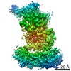
| Structure viewer | Molecule: MolmilJmol/JSmol |
|---|
- Downloads & links
-Download
| PDBx/mmCIF format | 1ujn.cif.gz | 150 KB | Display | PDBx/mmCIF format |
|---|---|---|---|---|
| PDB format | pdb1ujn.ent.gz | 117.7 KB | Display | PDB format |
| PDBx/mmJSON format | 1ujn.json.gz | Tree view | PDBx/mmJSON format | |
| Others |  Other downloads Other downloads |
-Validation report
| Arichive directory | https://data.pdbj.org/pub/pdb/validation_reports/uj/1ujnftp://data.pdbj.org/pub/pdb/validation_reports/uj/1ujn | HTTPS FTP |
|---|
-Related structure data
| Related structure data | 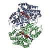 1dqsS S: Starting model for refinement |
|---|---|
| Similar structure data | |
| Other databases |


-Links
 PDBj
PDBj- Assembly
| Deposited unit | 
| ||||||||
|---|---|---|---|---|---|---|---|---|---|
| 1 |
| ||||||||
| Unit cell |
| ||||||||
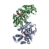
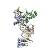
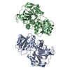
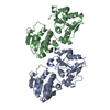
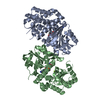
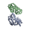
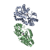
| Details | The biological assembly is a dimer in the asymmetric unit. |
-Components
| #1: Protein | Mass: 37559.043 Da / Num. of mol.: 2 Source method: isolated from a genetically manipulated source Source: (gene. exp.) Thermus thermophilus (bacteria) / Strain: HB8 / Plasmid: pET11a / Production host: #2: Water | ChemComp-HOH / |  Mass: 18.015 Da / Num. of mol.: 645 / Source method: isolated from a natural source / Formula: H2O Mass: 18.015 Da / Num. of mol.: 645 / Source method: isolated from a natural source / Formula: H2O |
|---|
-Experimental details
-Experiment
| Experiment | Method: X-RAY DIFFRACTION / Number of used crystals: 1 |
|---|
- Sample preparation
| Crystal | Density Matthews: 1.99 Å3/Da / Density % sol: 37.79 % | ||||||||||||||||||||||||
|---|---|---|---|---|---|---|---|---|---|---|---|---|---|---|---|---|---|---|---|---|---|---|---|---|---|
| Crystal grow | Temperature: 291 K / Method: microbatch / pH: 5.6 / Details: PEG 4000, pH 5.6, MICROBATCH, temperature 291K | ||||||||||||||||||||||||
| Crystal grow | *PLUS Temperature: 18 ℃ / Method: batch method | ||||||||||||||||||||||||
| Components of the solutions | *PLUS
|
-Data collection
| Diffraction | Mean temperature: 100 K |
|---|---|
| Diffraction source | Source: SYNCHROTRON / Site: SPring-8  / Beamline: BL26B1 / Wavelength: 1 Å / Beamline: BL26B1 / Wavelength: 1 Å |
| Detector | Type: RIGAKU RAXIS V / Detector: IMAGE PLATE / Date: Nov 28, 2002 / Details: mirrors |
| Radiation | Monochromator: mirrors / Protocol: SINGLE WAVELENGTH / Monochromatic (M) / Laue (L): M / Scattering type: x-ray |
| Radiation wavelength | Wavelength: 1 Å / Relative weight: 1 |
| Reflection | Resolution: 1.8→50 Å / Num. all: 58577 / Num. obs: 58577 / % possible obs: 99.4 % / Observed criterion σ(F): 0 / Observed criterion σ(I): 0 / Redundancy: 3.71 % / Biso Wilson estimate: 26.9 Å2 / Rmerge(I) obs: 0.04 / Rsym value: 0.035 / Net I/σ(I): 16.2 |
| Reflection shell | Resolution: 1.8→1.86 Å / Redundancy: 3.72 % / Rmerge(I) obs: 0.504 / Mean I/σ(I) obs: 3.35 / Num. unique all: 5792 / Rsym value: 0.417 / % possible all: 99 |
| Reflection | *PLUS Num. measured all: 217026 / Rmerge(I) obs: 0.04 |
| Reflection shell | *PLUS % possible obs: 99 % / Mean I/σ(I) obs: 3.4 |
- Processing
| Software |
| |||||||||||||||||||||||||
|---|---|---|---|---|---|---|---|---|---|---|---|---|---|---|---|---|---|---|---|---|---|---|---|---|---|---|
| Refinement | Method to determine structure: MOLECULAR REPLACEMENT Starting model: 1DQS Resolution: 1.8→37.2 Å / Isotropic thermal model: Anisotropic / Cross valid method: THROUGHOUT / σ(F): 0 / Stereochemistry target values: Engh & Huber
| |||||||||||||||||||||||||
| Displacement parameters | Biso mean: 33.5 Å2
| |||||||||||||||||||||||||
| Refine analyze |
| |||||||||||||||||||||||||
| Refinement step | Cycle: LAST / Resolution: 1.8→37.2 Å
| |||||||||||||||||||||||||
| Refine LS restraints |
| |||||||||||||||||||||||||
| LS refinement shell | Resolution: 1.8→1.86 Å / Rfactor Rfree error: 0.017
| |||||||||||||||||||||||||
| Refinement | *PLUS Lowest resolution: 40 Å / Rfactor Rfree: 0.24 / Rfactor Rwork: 0.2 | |||||||||||||||||||||||||
| Solvent computation | *PLUS | |||||||||||||||||||||||||
| Displacement parameters | *PLUS | |||||||||||||||||||||||||
| Refine LS restraints | *PLUS
|
