 ムービー
ムービー コントローラー


+ データを開く

- 基本情報
基本情報
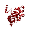
| 登録情報 | データベース: PDB / ID: 1u98 | ||||||
|---|---|---|---|---|---|---|---|
| タイトル | Crystal Structure of E. coli RecA in a Compressed Helical Filament Form3 | ||||||
 要素 要素 | RecA protein | ||||||
 キーワード キーワード | DNA BINDING PROTEIN / RecA / homologous recombination / ATPase / DNA repair | ||||||
| 機能・相同性 |  機能・相同性情報 機能・相同性情報DNA polymerase V complex / homologous recombination / SOS response / recombinational repair / response to ionizing radiation / ATP-dependent DNA damage sensor activity / ATP-dependent activity, acting on DNA / translesion synthesis / cell motility / single-stranded DNA binding ...DNA polymerase V complex / homologous recombination / SOS response / recombinational repair / response to ionizing radiation / ATP-dependent DNA damage sensor activity / ATP-dependent activity, acting on DNA / translesion synthesis / cell motility / single-stranded DNA binding / DNA recombination / DNA-binding transcription factor binding / damaged DNA binding / DNA damage response / ATP hydrolysis activity / ATP binding / cytoplasm 類似検索 - 分子機能 | ||||||
| 生物種 |  | ||||||
| 手法 |  X線回折 / シンクロトロン / 分子置換 / 解像度: 2 Å X線回折 / シンクロトロン / 分子置換 / 解像度: 2 Å | ||||||
 データ登録者 データ登録者 | Xing, X. / Bell, C.E. | ||||||
 引用 引用 | ジャーナル: J.Mol.Biol. / 年: 2004 タイトル: Crystal structures of Escherichia coli RecA in a compressed helical filament. 著者: Xing, X. / Bell, C.E. | ||||||
| 履歴 |
|
- 構造の表示
構造の表示
| 構造ビューア | 分子: MolmilJmol/JSmol |
|---|
- ダウンロードとリンク
-ダウンロード
| PDBx/mmCIF形式 | 1u98.cif.gz | 78.4 KB | 表示 | PDBx/mmCIF形式 |
|---|---|---|---|---|
| PDB形式 | pdb1u98.ent.gz | 56.1 KB | 表示 | PDB形式 |
| PDBx/mmJSON形式 | 1u98.json.gz | ツリー表示 | PDBx/mmJSON形式 | |
| その他 |  その他のダウンロード その他のダウンロード |
-検証レポート
| 文書・要旨 | 1u98_validation.pdf.gz | 444.3 KB | 表示 | wwPDB検証レポート |
|---|---|---|---|---|
| 文書・詳細版 | 1u98_full_validation.pdf.gz | 448.3 KB | 表示 | |
| XML形式データ | 1u98_validation.xml.gz | 15.2 KB | 表示 | |
| CIF形式データ | 1u98_validation.cif.gz | 21.4 KB | 表示 | |
| アーカイブディレクトリ | https://data.pdbj.org/pub/pdb/validation_reports/u9/1u98ftp://data.pdbj.org/pub/pdb/validation_reports/u9/1u98 | HTTPS FTP |
-関連構造データ


-リンク
 PDBj
PDBj
- 集合体
集合体
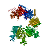
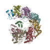
| 登録構造単位 | 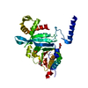
| ||||||||
|---|---|---|---|---|---|---|---|---|---|
| 1 | x 6
| ||||||||
| 単位格子 |
| ||||||||
| 詳細 | The biological assembly is a continuous helical filament formed by the 6sub1 symmetry operators |
-要素
| #1: タンパク質 | 分子量: 38298.559 Da / 分子数: 1 / 由来タイプ: 組換発現 / 詳細: RECA WITH N-TERMINAL GSHM RESIDUES / 由来: (組換発現) | ||
|---|---|---|---|
| #2: 化合物 | ChemComp-SO4 /   分子量: 96.063 Da / 分子数: 1 / 由来タイプ: 合成 / 式: SO4 分子量: 96.063 Da / 分子数: 1 / 由来タイプ: 合成 / 式: SO4 | ||
| #3: 化合物 |   分子量: 92.094 Da / 分子数: 2 / 由来タイプ: 合成 / 式: C3H8O3 分子量: 92.094 Da / 分子数: 2 / 由来タイプ: 合成 / 式: C3H8O3#4: 水 | ChemComp-HOH / |  分子量: 18.015 Da / 分子数: 172 / 由来タイプ: 天然 / 式: H2O 分子量: 18.015 Da / 分子数: 172 / 由来タイプ: 天然 / 式: H2O |
-実験情報
-実験
| 実験 | 手法: X線回折 / 使用した結晶の数: 1 |
|---|
- 試料調製
試料調製
| 結晶 | マシュー密度: 3.3 Å3/Da / 溶媒含有率: 63 % |
|---|---|
| 結晶化 | 温度: 295 K / 手法: 蒸気拡散法, ハンギングドロップ法 / pH: 5.4 詳細: PEG 2000 Monomethylether, lithium sulfate, sodium phosphate, sodium citrate, pH 5.4, VAPOR DIFFUSION, HANGING DROP, temperature 295K |
-データ収集
| 回折 | 平均測定温度: 100 K |
|---|---|
| 放射光源 | 由来: シンクロトロン / サイト: APS  / ビームライン: 19-ID / ビームライン: 19-ID |
| 検出器 | タイプ: SBC-2 / 検出器: CCD |
| 放射 | プロトコル: SINGLE WAVELENGTH / 単色(M)・ラウエ(L): M / 散乱光タイプ: x-ray |
| 放射波長 | 相対比: 1 |
| 反射 | 解像度: 2→31.29 Å / Num. all: 33414 / Num. obs: 33112 / % possible obs: 98.8 % / Observed criterion σ(F): 0 / Observed criterion σ(I): 0 / 冗長度: 11.8 % / Biso Wilson estimate: 26.7 Å2 / Limit h max: 46 / Limit h min: 1 / Limit k max: 46 / Limit k min: 1 / Limit l max: 36 / Limit l min: 0 / Observed criterion F max: 1093613.19 / Observed criterion F min: 0.32 / Rmerge(I) obs: 0.06 / Net I/σ(I): 63.3 |
| 反射 シェル | 解像度: 2→2.07 Å / Rmerge(I) obs: 0.735 / Mean I/σ(I) obs: 4.9 / % possible all: 99.5 |
- 解析
解析
| ソフトウェア |
| ||||||||||||||||||||||||||||||||||||||||||||||||||||||||||||||||||||||||||||||||||||||||||
|---|---|---|---|---|---|---|---|---|---|---|---|---|---|---|---|---|---|---|---|---|---|---|---|---|---|---|---|---|---|---|---|---|---|---|---|---|---|---|---|---|---|---|---|---|---|---|---|---|---|---|---|---|---|---|---|---|---|---|---|---|---|---|---|---|---|---|---|---|---|---|---|---|---|---|---|---|---|---|---|---|---|---|---|---|---|---|---|---|---|---|---|
| 精密化 | 構造決定の手法: 分子置換 開始モデル: PDB ID 2REB 解像度: 2→31.29 Å / Rfactor Rfree error: 0.004 / Occupancy max: 1 / Occupancy min: 1 / 交差検証法: THROUGHOUT / σ(F): 0 / 立体化学のターゲット値: Engh & Huber
| ||||||||||||||||||||||||||||||||||||||||||||||||||||||||||||||||||||||||||||||||||||||||||
| 溶媒の処理 | 溶媒モデル: CNS bulk solvent model used / Bsol: 72.6877 Å2 / ksol: 0.403562 e/Å3 | ||||||||||||||||||||||||||||||||||||||||||||||||||||||||||||||||||||||||||||||||||||||||||
| 原子変位パラメータ | Biso max: 119.57 Å2 / Biso mean: 50.41 Å2 / Biso min: 20.92 Å2
| ||||||||||||||||||||||||||||||||||||||||||||||||||||||||||||||||||||||||||||||||||||||||||
| Refine Biso |
| ||||||||||||||||||||||||||||||||||||||||||||||||||||||||||||||||||||||||||||||||||||||||||
| Refine analyze |
| ||||||||||||||||||||||||||||||||||||||||||||||||||||||||||||||||||||||||||||||||||||||||||
| 精密化ステップ | サイクル: LAST / 解像度: 2→31.29 Å
| ||||||||||||||||||||||||||||||||||||||||||||||||||||||||||||||||||||||||||||||||||||||||||
| 拘束条件 |
| ||||||||||||||||||||||||||||||||||||||||||||||||||||||||||||||||||||||||||||||||||||||||||
| LS精密化 シェル | Refine-ID: X-RAY DIFFRACTION
| ||||||||||||||||||||||||||||||||||||||||||||||||||||||||||||||||||||||||||||||||||||||||||
| Xplor file |
|
