 Movie
Movie Controller


+ Open data

- Basic information
| Entry | Database: PDB / ID: 2reb | ||||||
|---|---|---|---|---|---|---|---|
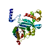
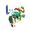
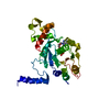
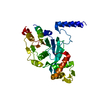
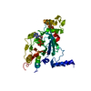
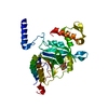
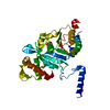
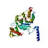
| Title | THE STRUCTURE OF THE E. COLI RECA PROTEIN MONOMER AND POLYMER | ||||||
 Components Components | REC A | ||||||
 Keywords Keywords | DNA BINDING PROTEIN / SELF-CLEAVAGE STIMULATION / HOMOLOGOUS RECOMBINATION | ||||||
| Function / homology |  Function and homology information Function and homology informationDNA polymerase V complex / homologous recombination / SOS response / recombinational repair / ATP-dependent DNA damage sensor activity / response to ionizing radiation / ATP-dependent activity, acting on DNA / translesion synthesis / cell motility / single-stranded DNA binding ...DNA polymerase V complex / homologous recombination / SOS response / recombinational repair / ATP-dependent DNA damage sensor activity / response to ionizing radiation / ATP-dependent activity, acting on DNA / translesion synthesis / cell motility / single-stranded DNA binding / DNA recombination / DNA-binding transcription factor binding / damaged DNA binding / DNA damage response / ATP binding / cytoplasm Similarity search - Function | ||||||
| Biological species |  | ||||||
| Method |  X-RAY DIFFRACTION / Resolution: 2.3 Å X-RAY DIFFRACTION / Resolution: 2.3 Å | ||||||
 Authors Authors | Story, R.M. / Steitz, T.A. | ||||||
 Citation Citation | Journal: Nature / Year: 1992 Title: The structure of the E. coli recA protein monomer and polymer. Authors: Story, R.M. / Weber, I.T. / Steitz, T.A. #1: Journal: Nature / Year: 1992Title: Structure of the Reca Protein-Adp Complex Authors: Story, R.M. / Steitz, T.A. #2: Journal: Nature / Year: 1992Title: The Structure of the E. Coli Reca Protein Monomer and Polymer: Erratum Authors: Story, R.M. / Weber, I.T. / Steitz, T.A. | ||||||
| History |
|
- Structure visualization
| Structure viewer | Molecule: MolmilJmol/JSmol |
|---|
- Downloads & links
-Download
| PDBx/mmCIF format | 2reb.cif.gz | 70.9 KB | Display | PDBx/mmCIF format |
|---|---|---|---|---|
| PDB format | pdb2reb.ent.gz | 52.7 KB | Display | PDB format |
| PDBx/mmJSON format | 2reb.json.gz | Tree view | PDBx/mmJSON format | |
| Others |  Other downloads Other downloads |
-Validation report
| Arichive directory | https://data.pdbj.org/pub/pdb/validation_reports/re/2rebftp://data.pdbj.org/pub/pdb/validation_reports/re/2reb | HTTPS FTP |
|---|
-Related structure data
| Similar structure data |
|---|


-Links
 PDBj
PDBj
- Assembly
| Deposited unit | 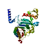
| ||||||||
|---|---|---|---|---|---|---|---|---|---|
| 1 |
| ||||||||
| Unit cell |
| ||||||||
| Atom site foot note | 1: THE PEPTIDE BOND BETWEEN ASP 144 AND SER 145 HAS BEEN BUILT IN THE CIS CONFORMATION. |
-Components
| #1: Protein | Mass: 37885.086 Da / Num. of mol.: 1 Source method: isolated from a genetically manipulated source Source: (gene. exp.) |
|---|---|
| #2: Water | ChemComp-HOH /  Mass: 18.015 Da / Num. of mol.: 136 / Source method: isolated from a natural source / Formula: H2O Mass: 18.015 Da / Num. of mol.: 136 / Source method: isolated from a natural source / Formula: H2O |
-Experimental details
-Experiment
| Experiment | Method: X-RAY DIFFRACTION |
|---|
- Sample preparation
| Crystal | Density Matthews: 3.3 Å3/Da / Density % sol: 62.76 % | |||||||||||||||||||||||||||||||||||||||||||||||||||||||||||||||
|---|---|---|---|---|---|---|---|---|---|---|---|---|---|---|---|---|---|---|---|---|---|---|---|---|---|---|---|---|---|---|---|---|---|---|---|---|---|---|---|---|---|---|---|---|---|---|---|---|---|---|---|---|---|---|---|---|---|---|---|---|---|---|---|---|
| Crystal grow | *PLUS pH: 5.4 / Method: microdialysis | |||||||||||||||||||||||||||||||||||||||||||||||||||||||||||||||
| Components of the solutions | *PLUS
|
-Data collection
| Radiation | Scattering type: x-ray |
|---|---|
| Radiation wavelength | Relative weight: 1 |
| Reflection | *PLUS Highest resolution: 2.3 Å / % possible obs: 92 % / Rmerge(I) obs: 0.055 |
- Processing
| Software |
| ||||||||||||||||||||||||||||||||||||||||||||||||||||||||||||
|---|---|---|---|---|---|---|---|---|---|---|---|---|---|---|---|---|---|---|---|---|---|---|---|---|---|---|---|---|---|---|---|---|---|---|---|---|---|---|---|---|---|---|---|---|---|---|---|---|---|---|---|---|---|---|---|---|---|---|---|---|---|
| Refinement | Rfactor Rwork: 0.21 / Rfactor obs: 0.21 / Highest resolution: 2.3 Å | ||||||||||||||||||||||||||||||||||||||||||||||||||||||||||||
| Refinement step | Cycle: LAST / Highest resolution: 2.3 Å
| ||||||||||||||||||||||||||||||||||||||||||||||||||||||||||||
| Refine LS restraints |
|
