 Movie
Movie Controller

[English] 日本語
 Yorodumi
- PDB-1u7z: Phosphopantothenoylcysteine synthetase from E. coli, 4'-phosphopa... -
+ Open data

- Basic information
| Entry | Database: PDB / ID: 1u7z | ||||||
|---|---|---|---|---|---|---|---|
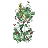
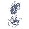
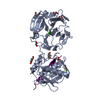
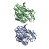
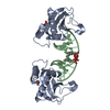
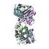
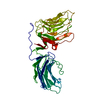
| Title | Phosphopantothenoylcysteine synthetase from E. coli, 4'-phosphopantothenoyl-CMP complex | ||||||
 Components Components | Coenzyme A biosynthesis bifunctional protein coaBC | ||||||
 Keywords Keywords | LIGASE / Coenzyme A biosynthesis | ||||||
| Function / homology |  Function and homology information Function and homology informationpantothenate catabolic process / phosphopantothenoylcysteine decarboxylase / phosphopantothenate-cysteine ligase (CTP) / phosphopantothenate--cysteine ligase activity / phosphopantothenoylcysteine decarboxylase complex / phosphopantothenoylcysteine decarboxylase activity / coenzyme A biosynthetic process / FMN binding / metal ion binding / identical protein binding / cytosol Similarity search - Function | ||||||
| Biological species |  | ||||||
| Method |  X-RAY DIFFRACTION / SYNCHROTRON / MOLECULAR REPLACEMENT / Resolution: 2.3 Å X-RAY DIFFRACTION / SYNCHROTRON / MOLECULAR REPLACEMENT / Resolution: 2.3 Å | ||||||
 Authors Authors | Stanitzek, S. / Augustin, M.A. / Huber, R. / Kupke, T. / Steinbacher, S. | ||||||
 Citation Citation | Journal: STRUCTURE / Year: 2004 Title: Structural Basis of CTP-Dependent Peptide Bond Formation in Coenzyme A Biosynthesis Catalyzed by Escherichia coli PPC Synthetase Authors: Stanitzek, S. / Augustin, M.A. / Huber, R. / Kupke, T. / Steinbacher, S. | ||||||
| History |
|
- Structure visualization
| Structure viewer | Molecule: MolmilJmol/JSmol |
|---|
- Downloads & links
-Download
| PDBx/mmCIF format | 1u7z.cif.gz | 145.2 KB | Display | PDBx/mmCIF format |
|---|---|---|---|---|
| PDB format | pdb1u7z.ent.gz | 114.4 KB | Display | PDB format |
| PDBx/mmJSON format | 1u7z.json.gz | Tree view | PDBx/mmJSON format | |
| Others |  Other downloads Other downloads |
-Validation report
| Summary document | 1u7z_validation.pdf.gz | 1.2 MB | Display | wwPDB validaton report |
|---|---|---|---|---|
| Full document | 1u7z_full_validation.pdf.gz | 1.2 MB | Display | |
| Data in XML | 1u7z_validation.xml.gz | 30 KB | Display | |
| Data in CIF | 1u7z_validation.cif.gz | 41.3 KB | Display | |
| Arichive directory | https://data.pdbj.org/pub/pdb/validation_reports/u7/1u7zftp://data.pdbj.org/pub/pdb/validation_reports/u7/1u7z | HTTPS FTP |
-Related structure data
| Related structure data | 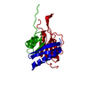 1u7uSC 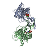 1u7wC 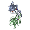 1u80C S: Starting model for refinement C: citing same article ( |
|---|---|
| Similar structure data |


-Links
 PDBj
PDBj- Assembly
| Deposited unit | 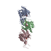
| ||||||||
|---|---|---|---|---|---|---|---|---|---|
| 1 | 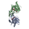
| ||||||||
| 2 | 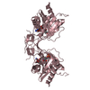
| ||||||||
| Unit cell |
|
-Components
| #1: Protein | Mass: 24833.348 Da / Num. of mol.: 3 Fragment: Phosphopantothenoylcysteine synthetase(residues 181-406) Mutation: N210D Source method: isolated from a genetically manipulated source Source: (gene. exp.) References: UniProt: P0ABQ0, phosphopantothenate-cysteine ligase (CTP) #2: Chemical |   Type: RNA linking / Mass: 604.396 Da / Num. of mol.: 3 / Source method: obtained synthetically / Formula: C18H30N4O15P2 Type: RNA linking / Mass: 604.396 Da / Num. of mol.: 3 / Source method: obtained synthetically / Formula: C18H30N4O15P2#3: Water | ChemComp-HOH / |  Mass: 18.015 Da / Num. of mol.: 258 / Source method: isolated from a natural source / Formula: H2O Mass: 18.015 Da / Num. of mol.: 258 / Source method: isolated from a natural source / Formula: H2O |
|---|
-Experimental details
-Experiment
| Experiment | Method: X-RAY DIFFRACTION / Number of used crystals: 1 |
|---|
- Sample preparation
| Crystal | Density Matthews: 2.55 Å3/Da / Density % sol: 51.82 % |
|---|---|
| Crystal grow | Temperature: 293 K / Method: vapor diffusion, sitting drop / pH: 6.6 Details: PEG 3000, pH 6.6, VAPOR DIFFUSION, SITTING DROP, temperature 293K |
-Data collection
| Diffraction | Mean temperature: 100 K |
|---|---|
| Diffraction source | Source: SYNCHROTRON / Site: MPG/DESY, HAMBURG  / Beamline: BW6 / Wavelength: 0.9789 Å / Beamline: BW6 / Wavelength: 0.9789 Å |
| Detector | Type: MARRESEARCH / Detector: CCD / Date: Nov 12, 2003 |
| Radiation | Protocol: SINGLE WAVELENGTH / Monochromatic (M) / Laue (L): M / Scattering type: x-ray |
| Radiation wavelength | Wavelength: 0.9789 Å / Relative weight: 1 |
| Reflection | Resolution: 2.3→20 Å / Num. all: 32347 / Num. obs: 32347 / % possible obs: 93.4 % / Observed criterion σ(F): 0 / Observed criterion σ(I): 0 / Redundancy: 2.9 % / Rmerge(I) obs: 0.088 |
| Reflection shell | Resolution: 2.3→2.38 Å / Rmerge(I) obs: 0.297 / % possible all: 97.1 |
- Processing
| Software |
| |||||||||||||||||||||||||
|---|---|---|---|---|---|---|---|---|---|---|---|---|---|---|---|---|---|---|---|---|---|---|---|---|---|---|
| Refinement | Method to determine structure: MOLECULAR REPLACEMENT Starting model: 1U7U Resolution: 2.3→20 Å / σ(F): 0 / Stereochemistry target values: Engh & Huber
| |||||||||||||||||||||||||
| Refinement step | Cycle: LAST / Resolution: 2.3→20 Å
| |||||||||||||||||||||||||
| Refine LS restraints |
|
