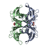
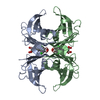
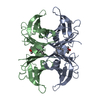
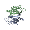
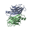
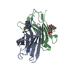
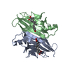
SEQUENCE Three of the four monomers in the transthyretin tetramer contained a C10A mutation. The ... SEQUENCE Three of the four monomers in the transthyretin tetramer contained a C10A mutation. The fourth monomer was wildtype, with a Cys at the 10 position and was chemically linked via a disulfide bridge to the P2C small molecule. However due to crystal symmetry and the statistically random way in which the tetramers were added to the crystal, there is no way tO distinguish the subunits apart. Therefore each chain in the asymmetric unit contains the wild type sequence. The C10A mutant monomer has DYKDDDYKDYKDDDYK added to the N-terminus and the vector used was the plamid PMMHa. See the paper for details.
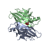
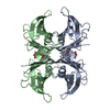
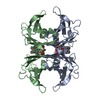
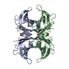
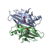
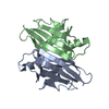
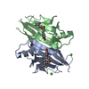
The biological assembly is a teteramer generated from the dimer in the asymmetric unit by the operation: 1 0 0 0 0 1 0 0 0 0 1 0 -1 0 0 43.65 0 -1 0 86.63 0 0 1 0
-
Components
#1: Protein
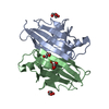
Transthyretin / Prealbumin / TBPA / TTR / ATTR
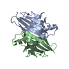
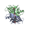
Mass: 13777.360 Da / Num. of mol.: 2 Source method: isolated from a genetically manipulated source Details: see remark 999 for mutation information / Source: (gene. exp.) Homo sapiens (human) / Gene: TTR; PALB; / Plasmid: pET2c / Production host: Escherichia coli (E. coli) / Strain (production host): BL21 (DE3) Epicurian Gold / References: UniProt: P02766
Resolution: 1.69→27.63 Å / Cor.coef. Fo:Fc: 0.947 / Cor.coef. Fo:Fc free: 0.937 / SU B: 2.602 / SU ML: 0.084 / TLS residual ADP flag: LIKELY RESIDUAL / Cross valid method: THROUGHOUT / ESU R: 0.126 / ESU R Free: 0.115 / Stereochemistry target values: MAXIMUM LIKELIHOOD Details: HYDROGENS HAVE BEEN ADDED IN THE RIDING POSITIONS, SEE REMARK 999 FOR MUTATIONS AND FURTHER REFINEMENT INFORMATION
Rfactor
Num. reflection
% reflection
Selection details
Rfree
0.23865
1283
4.8 %
RANDOM
Rwork
0.21676
-
-
-
obs
0.21783
25202
96.04 %
-
all
-
27493
-
-
Solvent computation
Ion probe radii: 0.8 Å / Shrinkage radii: 0.8 Å / VDW probe radii: 1.4 Å / Solvent model: BABINET MODEL WITH MASK
Displacement parameters
Biso mean: 15.538 Å2
Baniso -1
Baniso -2
Baniso -3
1-
0.24 Å2
0 Å2
0 Å2
2-
-
-0.22 Å2
0 Å2
3-
-
-
-0.02 Å2
Refinement step
Cycle: LAST / Resolution: 1.69→27.63 Å
Protein
Nucleic acid
Ligand
Solvent
Total
Num. atoms
1792
0
42
104
1938
Refine LS restraints
Refine-ID
Type
Dev ideal
Dev ideal target
Number
X-RAY DIFFRACTION
r_bond_refined_d
0.015
0.021
1886
X-RAY DIFFRACTION
r_bond_other_d
0.002
0.02
1644
X-RAY DIFFRACTION
r_angle_refined_deg
1.552
1.963
2574
X-RAY DIFFRACTION
r_angle_other_deg
0.759
3
3824
X-RAY DIFFRACTION
r_dihedral_angle_1_deg
6.175
5
230
X-RAY DIFFRACTION
r_chiral_restr
0.086
0.2
286
X-RAY DIFFRACTION
r_gen_planes_refined
0.009
0.02
2104
X-RAY DIFFRACTION
r_gen_planes_other
0.012
0.02
392
X-RAY DIFFRACTION
r_nbd_refined
0.212
0.3
298
X-RAY DIFFRACTION
r_nbd_other
0.276
0.3
1835
X-RAY DIFFRACTION
r_nbtor_other
0.094
0.5
1167
X-RAY DIFFRACTION
r_xyhbond_nbd_refined
0.196
0.5
181
X-RAY DIFFRACTION
r_symmetry_vdw_refined
0.223
0.3
30
X-RAY DIFFRACTION
r_symmetry_vdw_other
0.278
0.3
95
X-RAY DIFFRACTION
r_symmetry_hbond_refined
0.168
0.5
23
X-RAY DIFFRACTION
r_mcbond_it
0.895
1.5
1160
X-RAY DIFFRACTION
r_mcangle_it
1.681
2
1882
X-RAY DIFFRACTION
r_scbond_it
2.628
3
726
X-RAY DIFFRACTION
r_scangle_it
4.194
4.5
692
LS refinement shell
Resolution: 1.689→1.733 Å / Total num. of bins used: 20
Rfactor
Num. reflection
Rfree
0.363
87
Rwork
0.337
1534
obs
-
1534
Refinement TLS params.
Method: refined / Refine-ID: X-RAY DIFFRACTION
ID
L11 (°2)
L12 (°2)
L13 (°2)
L22 (°2)
L23 (°2)
L33 (°2)
S11 (Å °)
S12 (Å °)
S13 (Å °)
S21 (Å °)
S22 (Å °)
S23 (Å °)
S31 (Å °)
S32 (Å °)
S33 (Å °)
T11 (Å2)
T12 (Å2)
T13 (Å2)
T22 (Å2)
T23 (Å2)
T33 (Å2)
Origin x (Å)
Origin y (Å)
Origin z (Å)
1
3.2039
1.403
0.7625
2.0974
1.2524
1.4649
-0.0616
0.1417
-0.1437
-0.08
-0.0194
0.0533
0.0378
-0.0348
0.081
0.0037
0.0086
0.0145
0.1152
-0.015
0.0825
22.462
29.848
37.996
2
2.5525
-1.3792
0.788
2.7102
-0.9373
1.8909
-0.0134
-0.1537
-0.1992
0.2776
-0.0657
-0.0697
0.1314
0.0806
0.0791
0.0767
-0.0261
0.0178
0.1306
0.0247
0.0809
19.997
30.157
57.824
Refinement TLS group
ID
Refine-ID
Refine TLS-ID
Auth asym-ID
Label asym-ID
Auth seq-ID
Label seq-ID
1
X-RAY DIFFRACTION
1
A
A
10 - 125
10 - 125
2
X-RAY DIFFRACTION
2
B
B
10 - 125
10 - 125
+
About Yorodumi
-
News
-
Feb 9, 2022. New format data for meta-information of EMDB entries
New format data for meta-information of EMDB entries
Version 3 of the EMDB header file is now the official format.
The previous official version 1.9 will be removed from the archive.
In the structure databanks used in Yorodumi, some data are registered as the other names, "COVID-19 virus" and "2019-nCoV". Here are the details of the virus and the list of structure data.
Jan 31, 2019. EMDB accession codes are about to change! (news from PDBe EMDB page)
EMDB accession codes are about to change! (news from PDBe EMDB page)
The allocation of 4 digits for EMDB accession codes will soon come to an end. Whilst these codes will remain in use, new EMDB accession codes will include an additional digit and will expand incrementally as the available range of codes is exhausted. The current 4-digit format prefixed with “EMD-” (i.e. EMD-XXXX) will advance to a 5-digit format (i.e. EMD-XXXXX), and so on. It is currently estimated that the 4-digit codes will be depleted around Spring 2019, at which point the 5-digit format will come into force.
The EM Navigator/Yorodumi systems omit the EMD- prefix.
Related info.:Q: What is EMD? / ID/Accession-code notation in Yorodumi/EM Navigator
Yorodumi is a browser for structure data from EMDB, PDB, SASBDB, etc.
This page is also the successor to EM Navigator detail page, and also detail information page/front-end page for Omokage search.
The word "yorodu" (or yorozu) is an old Japanese word meaning "ten thousand". "mi" (miru) is to see.
Related info.:EMDB / PDB / SASBDB / Comparison of 3 databanks / Yorodumi Search / Aug 31, 2016. New EM Navigator & Yorodumi / Yorodumi Papers / Jmol/JSmol / Function and homology information / Changes in new EM Navigator and Yorodumi
 Movie
Movie Controller


 Open data
 Basic information
 Components
 Keywords
 Function and homology information
 Homo sapiens (human)
 X-RAY DIFFRACTION /
  Authors
 Citation
 Structure visualization
 Downloads & links
 Other downloads


 PDBj
PDBj


 Assembly
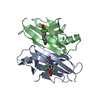


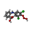
 Mass: 330.120 Da / Num. of mol.: 2 / Source method: obtained synthetically / Formula: C13H9Cl2NO5
 Mass: 18.015 Da / Num. of mol.: 104 / Source method: isolated from a natural source / Formula: H2O
 Sample preparation
 / Beamline: BL9-2 / Wavelength: 1.072 Å
 Processing