 Movie
Movie Controller

[English] 日本語
 Yorodumi
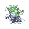
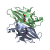
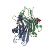
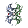
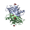
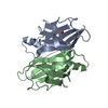
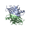
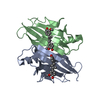
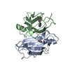
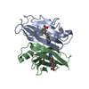
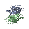
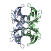
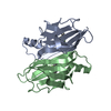
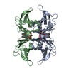
- PDB-2b15: The crystal structure of 2,4-dinitrophenol in complex with human ... -
+ Open data

- Basic information
| Entry | Database: PDB / ID: 2b15 | ||||||
|---|---|---|---|---|---|---|---|
| Title | The crystal structure of 2,4-dinitrophenol in complex with human transthyretin | ||||||
 Components Components | Transthyretin | ||||||
 Keywords Keywords | TRANSPORT PROTEIN / AMYLOID / TRANSTHYRETIN / 2 / 4-DINITROPHENOL / Tetramer Stabilizer | ||||||
| Function / homology |  Function and homology information Function and homology informationDefective visual phototransduction due to STRA6 loss of function / negative regulation of glomerular filtration / The canonical retinoid cycle in rods (twilight vision) / purine nucleobase metabolic process / hormone binding / Non-integrin membrane-ECM interactions / phototransduction, visible light / molecular sequestering activity / retinoid metabolic process / Retinoid metabolism and transport ...Defective visual phototransduction due to STRA6 loss of function / negative regulation of glomerular filtration / The canonical retinoid cycle in rods (twilight vision) / purine nucleobase metabolic process / hormone binding / Non-integrin membrane-ECM interactions / phototransduction, visible light / molecular sequestering activity / retinoid metabolic process / Retinoid metabolism and transport / hormone activity / azurophil granule lumen / Amyloid fiber formation / Neutrophil degranulation / protein-containing complex binding / protein-containing complex / : / extracellular exosome / extracellular region / identical protein binding Similarity search - Function | ||||||
| Biological species |  Homo sapiens (human) Homo sapiens (human) | ||||||
| Method |  X-RAY DIFFRACTION / SYNCHROTRON / MOLECULAR REPLACEMENT / Resolution: 1.7 Å X-RAY DIFFRACTION / SYNCHROTRON / MOLECULAR REPLACEMENT / Resolution: 1.7 Å | ||||||
 Authors Authors | Morais-de-Sa, E. / Neto-Silva, R.M. / Pereira, P.J. / Saraiva, M.J. / Damas, A.M. | ||||||
 Citation Citation | Journal: ACTA CRYSTALLOGR.,SECT.D / Year: 2006 Title: The binding of 2,4-dinitrophenol to wild-type and amyloidogenic transthyretin Authors: Morais-de-Sa, E. / Neto-Silva, R.M. / Pereira, P.J. / Saraiva, M.J. / Damas, A.M. | ||||||
| History |
|
- Structure visualization
| Structure viewer | Molecule: MolmilJmol/JSmol |
|---|
- Downloads & links
-Download
| PDBx/mmCIF format | 2b15.cif.gz | 60.3 KB | Display | PDBx/mmCIF format |
|---|---|---|---|---|
| PDB format | pdb2b15.ent.gz | 43.5 KB | Display | PDB format |
| PDBx/mmJSON format | 2b15.json.gz | Tree view | PDBx/mmJSON format | |
| Others |  Other downloads Other downloads |
-Validation report
| Arichive directory | https://data.pdbj.org/pub/pdb/validation_reports/b1/2b15ftp://data.pdbj.org/pub/pdb/validation_reports/b1/2b15 | HTTPS FTP |
|---|
-Related structure data
| Related structure data | 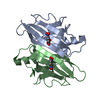 2b14C  2b16C  1f86S C: citing same article ( S: Starting model for refinement |
|---|---|
| Similar structure data |


-Links
 PDBj
PDBj


- Assembly
| Deposited unit | 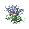
| ||||||||||||||||||||||||
|---|---|---|---|---|---|---|---|---|---|---|---|---|---|---|---|---|---|---|---|---|---|---|---|---|---|
| 1 | 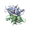
| ||||||||||||||||||||||||
| Unit cell |
| ||||||||||||||||||||||||
| Components on special symmetry positions |
|
-Components
| #1: Protein | Mass: 13777.360 Da / Num. of mol.: 2 Source method: isolated from a genetically manipulated source Source: (gene. exp.) Homo sapiens (human) / Plasmid: pINTR / Species (production host): Escherichia coli / Production host:  #2: Chemical |   Mass: 96.063 Da / Num. of mol.: 2 / Source method: obtained synthetically / Formula: SO4 Mass: 96.063 Da / Num. of mol.: 2 / Source method: obtained synthetically / Formula: SO4#3: Chemical | 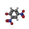  Mass: 184.106 Da / Num. of mol.: 2 / Source method: obtained synthetically / Formula: C6H4N2O5 Mass: 184.106 Da / Num. of mol.: 2 / Source method: obtained synthetically / Formula: C6H4N2O5#4: Water | ChemComp-HOH / |  Mass: 18.015 Da / Num. of mol.: 175 / Source method: isolated from a natural source / Formula: H2O Mass: 18.015 Da / Num. of mol.: 175 / Source method: isolated from a natural source / Formula: H2O |
|---|
-Experimental details
-Experiment
| Experiment | Method: X-RAY DIFFRACTION / Number of used crystals: 1 |
|---|
- Sample preparation
| Crystal | Density Matthews: 2.14 Å3/Da / Density % sol: 42.2 % |
|---|---|
| Crystal grow | Temperature: 287 K / Method: vapor diffusion, hanging drop / pH: 4.8 Details: ammonium sulfate, glycerol, sodium acetate, pH 4.8, VAPOR DIFFUSION, HANGING DROP, temperature 287K |
-Data collection
| Diffraction | Mean temperature: 100 K |
|---|---|
| Diffraction source | Source: SYNCHROTRON / Site: ESRF  / Beamline: ID14-3 / Wavelength: 0.934 Å / Beamline: ID14-3 / Wavelength: 0.934 Å |
| Detector | Type: ADSC QUANTUM 4 / Detector: CCD / Date: Jul 11, 2004 |
| Radiation | Protocol: SINGLE WAVELENGTH / Monochromatic (M) / Laue (L): M / Scattering type: x-ray |
| Radiation wavelength | Wavelength: 0.934 Å / Relative weight: 1 |
| Reflection | Resolution: 1.7→35.54 Å / Num. all: 25884 / Num. obs: 25884 / % possible obs: 98.5 % / Observed criterion σ(F): 0 / Observed criterion σ(I): 0 / Redundancy: 3.6 % / Rmerge(I) obs: 0.083 / Rsym value: 0.072 / Net I/σ(I): 5 |
| Reflection shell | Resolution: 1.7→1.79 Å / Redundancy: 3.6 % / Rmerge(I) obs: 0.212 / Mean I/σ(I) obs: 3.9 / Num. unique all: 3703 / Rsym value: 0.182 / % possible all: 98.6 |
- Processing
| Software |
| |||||||||||||||||||||||||
|---|---|---|---|---|---|---|---|---|---|---|---|---|---|---|---|---|---|---|---|---|---|---|---|---|---|---|
| Refinement | Method to determine structure: MOLECULAR REPLACEMENT Starting model: PDB ENTRY 1F86 Resolution: 1.7→10 Å / Cross valid method: THROUGHOUT / σ(F): 0 / Stereochemistry target values: Engh & Huber
| |||||||||||||||||||||||||
| Refinement step | Cycle: LAST / Resolution: 1.7→10 Å
| |||||||||||||||||||||||||
| Refine LS restraints |
|
