 Movie
Movie Controller

[English] 日本語
 Yorodumi
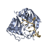
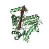
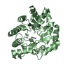
- PDB-1twu: 2.0 A Crystal Structure of a YycE Protein of Unknown Function fro... -
+ Open data

- Basic information
| Entry | Database: PDB / ID: 1twu | ||||||
|---|---|---|---|---|---|---|---|
| Title | 2.0 A Crystal Structure of a YycE Protein of Unknown Function from Bacillus subtilis, Putative Glyoxalase/Fosfomycin Resistance Protein | ||||||
 Components Components | Hypothetical protein yycE | ||||||
 Keywords Keywords | STRUCTURAL GENOMICS / UNKNOWN FUNCTION / Protein structure initiative / MCSG / duplication of the alpha-beta sandwichs. Bacillus subtilis / PSI / Midwest Center for Structural Genomics | ||||||
| Function / homology |  Function and homology information Function and homology information: / : / YycE-like N-terminal domain / YycE-like C-terminal domain / 2,3-Dihydroxybiphenyl 1,2-Dioxygenase, domain 1 / 2,3-Dihydroxybiphenyl 1,2-Dioxygenase; domain 1 / Glyoxalase/Bleomycin resistance protein/Dioxygenase superfamily / Vicinal oxygen chelate (VOC) domain / Vicinal oxygen chelate (VOC) domain profile. / Glyoxalase/Bleomycin resistance protein/Dihydroxybiphenyl dioxygenase ...: / : / YycE-like N-terminal domain / YycE-like C-terminal domain / 2,3-Dihydroxybiphenyl 1,2-Dioxygenase, domain 1 / 2,3-Dihydroxybiphenyl 1,2-Dioxygenase; domain 1 / Glyoxalase/Bleomycin resistance protein/Dioxygenase superfamily / Vicinal oxygen chelate (VOC) domain / Vicinal oxygen chelate (VOC) domain profile. / Glyoxalase/Bleomycin resistance protein/Dihydroxybiphenyl dioxygenase / Roll / Alpha Beta Similarity search - Domain/homology | ||||||
| Biological species |  | ||||||
| Method |  X-RAY DIFFRACTION / SYNCHROTRON / SAD / Resolution: 2 Å X-RAY DIFFRACTION / SYNCHROTRON / SAD / Resolution: 2 Å | ||||||
 Authors Authors | Zhang, R. / Quartey, P. / Collart, F. / Joachimiak, A. / Midwest Center for Structural Genomics (MCSG) | ||||||
 Citation Citation | Journal: To be Published Title: 2.0 A crystal structure of a hypothetical protein yycE from Bacillus subtilis Authors: Zhang, R. / Quartey, P. / Collart, F. / Joachimiak, A. | ||||||
| History |
|
- Structure visualization
| Structure viewer | Molecule: MolmilJmol/JSmol |
|---|
- Downloads & links
-Download
| PDBx/mmCIF format | 1twu.cif.gz | 40.3 KB | Display | PDBx/mmCIF format |
|---|---|---|---|---|
| PDB format | pdb1twu.ent.gz | 27.9 KB | Display | PDB format |
| PDBx/mmJSON format | 1twu.json.gz | Tree view | PDBx/mmJSON format | |
| Others |  Other downloads Other downloads |
-Validation report
| Arichive directory | https://data.pdbj.org/pub/pdb/validation_reports/tw/1twuftp://data.pdbj.org/pub/pdb/validation_reports/tw/1twu | HTTPS FTP |
|---|
-Related structure data
| Similar structure data | |
|---|---|
| Other databases |


-Links
 PDBj
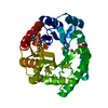
PDBj- Assembly
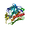
| Deposited unit | 
| ||||||||
|---|---|---|---|---|---|---|---|---|---|
| 1 | 
| ||||||||
| Unit cell |
| ||||||||
| Details | This protein exists as dimer.The second part of the biological assembly is generated by the two fold axis:-x,-y,-z |
-Components
| #1: Protein | Mass: 15604.514 Da / Num. of mol.: 1 Source method: isolated from a genetically manipulated source Source: (gene. exp.) |
|---|---|
| #2: Water | ChemComp-HOH /  Mass: 18.015 Da / Num. of mol.: 79 / Source method: isolated from a natural source / Formula: H2O Mass: 18.015 Da / Num. of mol.: 79 / Source method: isolated from a natural source / Formula: H2O |
-Experimental details
-Experiment
| Experiment | Method: X-RAY DIFFRACTION / Number of used crystals: 1 |
|---|
- Sample preparation
| Crystal | Density Matthews: 3.215 Å3/Da / Density % sol: 60.27 % |
|---|---|
| Crystal grow | Method: vapor diffusion, sitting drop / Details: VAPOR DIFFUSION, SITTING DROP |
-Data collection
| Diffraction | Mean temperature: 100 K |
|---|---|
| Diffraction source | Source: SYNCHROTRON / Site: APS  / Beamline: 19-BM / Wavelength: 0.9795 Å / Beamline: 19-BM / Wavelength: 0.9795 Å |
| Detector | Type: SBC-3 / Detector: CCD / Date: Jun 18, 2004 / Details: mirrors |
| Radiation | Monochromator: Si 111 channel / Protocol: SINGLE WAVELENGTH / Monochromatic (M) / Laue (L): M / Scattering type: x-ray |
| Radiation wavelength | Wavelength: 0.9795 Å / Relative weight: 1 |
| Reflection | Resolution: 2→50 Å / Num. all: 26277 / Num. obs: 25357 / % possible obs: 96.5 % / Observed criterion σ(F): 4 / Observed criterion σ(I): 4 / Redundancy: 5.2 % / Biso Wilson estimate: 16.4 Å2 / Rmerge(I) obs: 0.062 / Net I/σ(I): 23.7 |
| Reflection shell | Resolution: 2→2.07 Å / Redundancy: 3.6 % / Rmerge(I) obs: 0.287 / Mean I/σ(I) obs: 2.9 / Num. unique all: 2631 / % possible all: 75.6 |
- Processing
| Software |
| |||||||||||||||||||||||||
|---|---|---|---|---|---|---|---|---|---|---|---|---|---|---|---|---|---|---|---|---|---|---|---|---|---|---|
| Refinement | Method to determine structure: SAD / Resolution: 2→39.55 Å / Rfactor Rfree error: 0.007 / Data cutoff high absF: 269090.61 / Data cutoff low absF: 0 / Isotropic thermal model: RESTRAINED / Cross valid method: THROUGHOUT / σ(F): 0 / Stereochemistry target values: Engh & Huber
| |||||||||||||||||||||||||
| Solvent computation | Solvent model: FLAT MODEL / Bsol: 52.1179 Å2 / ksol: 0.361403 e/Å3 | |||||||||||||||||||||||||
| Displacement parameters | Biso mean: 33.9 Å2
| |||||||||||||||||||||||||
| Refine analyze |
| |||||||||||||||||||||||||
| Refinement step | Cycle: LAST / Resolution: 2→39.55 Å
| |||||||||||||||||||||||||
| Refine LS restraints |
| |||||||||||||||||||||||||
| LS refinement shell | Resolution: 2→2.13 Å / Rfactor Rfree error: 0.027 / Total num. of bins used: 6
| |||||||||||||||||||||||||
| Xplor file |
|
