 Movie
Movie Controller

[English] 日本語
 Yorodumi
- PDB-1tpy: Structure of the cyclopropane synthase MmaA2 from Mycobacterium t... -
+ Open data

- Basic information
| Entry | Database: PDB / ID: 1tpy | ||||||
|---|---|---|---|---|---|---|---|
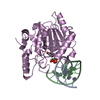
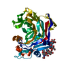
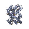
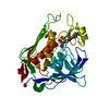
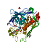
| Title | Structure of the cyclopropane synthase MmaA2 from Mycobacterium tuberculosis | ||||||
 Components Components | methoxy mycolic acid synthase 2 | ||||||
 Keywords Keywords | TRANSFERASE / Methyltransferase / cyclopropane synthase / mycolic acids / tuberculosis / SAM-dependent | ||||||
| Function / homology |  Function and homology information Function and homology informationcyclopropane-fatty-acyl-phospholipid synthase / cyclopropane-fatty-acyl-phospholipid synthase activity / mycolic acid biosynthetic process / lipid biosynthetic process / peptidoglycan-based cell wall / methyltransferase activity / methylation / plasma membrane Similarity search - Function | ||||||
| Biological species |   Mycobacterium tuberculosis (bacteria) Mycobacterium tuberculosis (bacteria) | ||||||
| Method |  X-RAY DIFFRACTION / SYNCHROTRON / MOLECULAR REPLACEMENT / Resolution: 2.2 Å X-RAY DIFFRACTION / SYNCHROTRON / MOLECULAR REPLACEMENT / Resolution: 2.2 Å | ||||||
 Authors Authors | Smith, C.V. / Sacchettini, J.C. | ||||||
 Citation Citation | Journal: To be Published Title: Structure of the cyclopropane synthase MmaA2 from Mycobacterium tuberculosis Authors: Smith, C.V. / Sacchettini, J.C. | ||||||
| History |
|
- Structure visualization
| Structure viewer | Molecule: MolmilJmol/JSmol |
|---|
- Downloads & links
-Download
| PDBx/mmCIF format | 1tpy.cif.gz | 75.7 KB | Display | PDBx/mmCIF format |
|---|---|---|---|---|
| PDB format | pdb1tpy.ent.gz | 54.9 KB | Display | PDB format |
| PDBx/mmJSON format | 1tpy.json.gz | Tree view | PDBx/mmJSON format | |
| Others |  Other downloads Other downloads |
-Validation report
| Arichive directory | https://data.pdbj.org/pub/pdb/validation_reports/tp/1tpyftp://data.pdbj.org/pub/pdb/validation_reports/tp/1tpy | HTTPS FTP |
|---|
-Related structure data
| Related structure data | 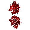 1lieS S: Starting model for refinement |
|---|---|
| Similar structure data |


-Links
 PDBj
PDBj


- Assembly
| Deposited unit | 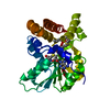
| ||||||||
|---|---|---|---|---|---|---|---|---|---|
| 1 |
| ||||||||
| Unit cell |
|
-Components
| #1: Protein | Mass: 32764.281 Da / Num. of mol.: 1 Source method: isolated from a genetically manipulated source Source: (gene. exp.) Mycobacterium tuberculosis (bacteria) / Gene: mmaA2 / Plasmid: pET30b-mmaA2 / Species (production host): Escherichia coli / Production host: References: UniProt: Q79FX6, cyclopropane-fatty-acyl-phospholipid synthase |
|---|---|
| #2: Chemical | ChemComp-CO3 /   Mass: 60.009 Da / Num. of mol.: 1 / Source method: obtained synthetically / Formula: CO3 Mass: 60.009 Da / Num. of mol.: 1 / Source method: obtained synthetically / Formula: CO3 |
| #3: Chemical | ChemComp-SAH /   Type: L-peptide linking / Mass: 384.411 Da / Num. of mol.: 1 / Source method: obtained synthetically / Formula: C14H20N6O5S Type: L-peptide linking / Mass: 384.411 Da / Num. of mol.: 1 / Source method: obtained synthetically / Formula: C14H20N6O5S |
| #4: Chemical | ChemComp-16A /   Mass: 284.543 Da / Num. of mol.: 1 / Source method: obtained synthetically / Formula: C19H42N Mass: 284.543 Da / Num. of mol.: 1 / Source method: obtained synthetically / Formula: C19H42N |
| #5: Water | ChemComp-HOH /  Mass: 18.015 Da / Num. of mol.: 192 / Source method: isolated from a natural source / Formula: H2O Mass: 18.015 Da / Num. of mol.: 192 / Source method: isolated from a natural source / Formula: H2O |
-Experimental details
-Experiment
| Experiment | Method: X-RAY DIFFRACTION / Number of used crystals: 1 |
|---|
- Sample preparation
| Crystal | Density Matthews: 2.541 Å3/Da / Density % sol: 49.73 % |
|---|---|
| Crystal grow | Temperature: 292 K / Method: vapor diffusion, sitting drop / pH: 6.5 Details: PEG 6000, MES buffer, cetyl trimethyl ammonium bromide, S-adenosyl-l-homocysteine, pH 6.5, VAPOR DIFFUSION, SITTING DROP, temperature 292K |
-Data collection
| Diffraction | Mean temperature: 100 K |
|---|---|
| Diffraction source | Source: SYNCHROTRON / Site: ALS  / Beamline: 5.0.3 / Wavelength: 1 Å / Beamline: 5.0.3 / Wavelength: 1 Å |
| Detector | Type: ADSC QUANTUM 4 / Detector: CCD / Date: Nov 15, 2002 |
| Radiation | Monochromator: Single crystal, cylindrically bent, Si(220) / Protocol: SINGLE WAVELENGTH / Monochromatic (M) / Laue (L): M / Scattering type: x-ray |
| Radiation wavelength | Wavelength: 1 Å / Relative weight: 1 |
| Reflection | Resolution: 2.2→50 Å / Num. all: 17966 / Num. obs: 17358 / % possible obs: 96.6 % / Observed criterion σ(F): 3 / Observed criterion σ(I): 3 / Redundancy: 3.9 % / Rmerge(I) obs: 0.0582 / Rsym value: 0.036 / Net I/σ(I): 10.8 |
| Reflection shell | Resolution: 2.19→2.3 Å / % possible all: 84.9 |
- Processing
| Software |
| |||||||||||||||||||||||||
|---|---|---|---|---|---|---|---|---|---|---|---|---|---|---|---|---|---|---|---|---|---|---|---|---|---|---|
| Refinement | Method to determine structure: MOLECULAR REPLACEMENT Starting model: PcaA ENTRY 1LIE Resolution: 2.2→50 Å / Cross valid method: THROUGHOUT / σ(F): 3 / Stereochemistry target values: Engh & Huber
| |||||||||||||||||||||||||
| Displacement parameters | Biso mean: 28.7 Å2 | |||||||||||||||||||||||||
| Refine analyze |
| |||||||||||||||||||||||||
| Refinement step | Cycle: LAST / Resolution: 2.2→50 Å
| |||||||||||||||||||||||||
| Refine LS restraints |
| |||||||||||||||||||||||||
| LS refinement shell | Resolution: 2.2→2.34 Å / Rfactor Rfree error: 0.016
|
