 Movie
Movie Controller

[English] 日本語
 Yorodumi
- PDB-1tp6: 1.5 A Crystal Structure of a NTF-2 Like Protein of Unknown Functi... -
+ Open data

- Basic information
| Entry | Database: PDB / ID: 1tp6 | ||||||
|---|---|---|---|---|---|---|---|
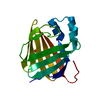
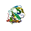
| Title | 1.5 A Crystal Structure of a NTF-2 Like Protein of Unknown Function PA1314 from Pseudomonas aeruginosa | ||||||
 Components Components | hypothetical protein PA1314 | ||||||
 Keywords Keywords | structural genomics / unknown function / Pseudomonas aeruginosa / alpha-beta sandwich / PSI / Protein Structure Initiative / Midwest Center for Structural Genomics / MCSG | ||||||
| Function / homology | Uncharacterised conserved protein UCP029394 / Domain of unknown function DUF4440 / Domain of unknown function (DUF4440) / Nuclear Transport Factor 2; Chain: A, - #50 / NTF2-like domain superfamily / Nuclear Transport Factor 2; Chain: A, / Roll / Alpha Beta / DUF4440 domain-containing protein Function and homology information Function and homology information | ||||||
| Biological species |  Pseudomonas aeruginosa PAO1 (bacteria) Pseudomonas aeruginosa PAO1 (bacteria) | ||||||
| Method |  X-RAY DIFFRACTION / SYNCHROTRON / MAD / Resolution: 1.5 Å X-RAY DIFFRACTION / SYNCHROTRON / MAD / Resolution: 1.5 Å | ||||||
 Authors Authors | Zhang, R. / Xu, L.X. / savchenko, A. / Edwards, A. / Joachimiak, A. / Midwest Center for Structural Genomics (MCSG) | ||||||
 Citation Citation | Journal: To be Published Title: 1.5A crystal structure of a hypothetical protein PA1314 from Pseudomonas aeruginosa Authors: Zhang, R. / Xu, L.X. / Savchenko, A. / Edwards, A. / Joachimiak, A. | ||||||
| History |
|
- Structure visualization
| Structure viewer | Molecule: MolmilJmol/JSmol |
|---|
- Downloads & links
-Download
| PDBx/mmCIF format | 1tp6.cif.gz | 38.8 KB | Display | PDBx/mmCIF format |
|---|---|---|---|---|
| PDB format | pdb1tp6.ent.gz | 26.3 KB | Display | PDB format |
| PDBx/mmJSON format | 1tp6.json.gz | Tree view | PDBx/mmJSON format | |
| Others |  Other downloads Other downloads |
-Validation report
| Summary document | 1tp6_validation.pdf.gz | 412.6 KB | Display | wwPDB validaton report |
|---|---|---|---|---|
| Full document | 1tp6_full_validation.pdf.gz | 413.6 KB | Display | |
| Data in XML | 1tp6_validation.xml.gz | 8.4 KB | Display | |
| Data in CIF | 1tp6_validation.cif.gz | 11.4 KB | Display | |
| Arichive directory | https://data.pdbj.org/pub/pdb/validation_reports/tp/1tp6ftp://data.pdbj.org/pub/pdb/validation_reports/tp/1tp6 | HTTPS FTP |
-Related structure data
| Similar structure data | |
|---|---|
| Other databases |


-Links
 PDBj
PDBj
- Assembly
| Deposited unit | 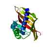
| ||||||||
|---|---|---|---|---|---|---|---|---|---|
| 1 |
| ||||||||
| Unit cell |
| ||||||||
| Details | This protein existed as monomer. The deposited coords. represents the monomer in the asymmtric unit. |
-Components
| #1: Protein | Mass: 14234.001 Da / Num. of mol.: 1 Source method: isolated from a genetically manipulated source Source: (gene. exp.) Pseudomonas aeruginosa PAO1 (bacteria) / Species: Pseudomonas aeruginosa / Strain: PA01 / Plasmid: pET15b / Species (production host): Escherichia coli / Production host: |
|---|---|
| #2: Water | ChemComp-HOH /  Mass: 18.015 Da / Num. of mol.: 143 / Source method: isolated from a natural source / Formula: H2O Mass: 18.015 Da / Num. of mol.: 143 / Source method: isolated from a natural source / Formula: H2O |
-Experimental details
-Experiment
| Experiment | Method: X-RAY DIFFRACTION / Number of used crystals: 1 |
|---|
- Sample preparation
| Crystal | Density Matthews: 1.741 Å3/Da / Density % sol: 27 % |
|---|---|
| Crystal grow | Temperature: 298 K / Method: vapor diffusion, hanging drop / pH: 4.2 Details: 0.1 M Sodium Acetate, 0.2 M Sodium Citrate, 4% Glycerol, 25% PEG 3350, pH 4.2, VAPOR DIFFUSION, HANGING DROP, temperature 298K |
-Data collection
| Diffraction | Mean temperature: 100 K | ||||||||||||
|---|---|---|---|---|---|---|---|---|---|---|---|---|---|
| Diffraction source | Source: SYNCHROTRON / Site: APS  / Beamline: 19-BM / Wavelength: 0.9795,0.9797,0.94656 / Beamline: 19-BM / Wavelength: 0.9795,0.9797,0.94656 | ||||||||||||
| Detector | Type: SBC-3 / Detector: CCD / Date: May 29, 2004 / Details: mirrors | ||||||||||||
| Radiation | Monochromator: Si 111 channel / Protocol: MAD / Monochromatic (M) / Laue (L): M / Scattering type: x-ray | ||||||||||||
| Radiation wavelength |
| ||||||||||||
| Reflection | Resolution: 1.5→50 Å / Num. all: 31652 / Num. obs: 30861 / % possible obs: 97.5 % / Observed criterion σ(F): 2 / Observed criterion σ(I): 4 / Redundancy: 4.4 % / Biso Wilson estimate: 13.6 Å2 / Rmerge(I) obs: 0.043 / Net I/σ(I): 35.07 | ||||||||||||
| Reflection shell | Resolution: 1.5→1.55 Å / Redundancy: 2.7 % / Rmerge(I) obs: 0.252 / Mean I/σ(I) obs: 3.8 / Num. unique all: 3202 / % possible all: 80.7 |
- Processing
| Software |
| |||||||||||||||||||||||||
|---|---|---|---|---|---|---|---|---|---|---|---|---|---|---|---|---|---|---|---|---|---|---|---|---|---|---|
| Refinement | Method to determine structure: MAD / Resolution: 1.5→29.06 Å / Isotropic thermal model: restrained / Cross valid method: THROUGHOUT / σ(F): 0 / Stereochemistry target values: Engh & Huber
| |||||||||||||||||||||||||
| Displacement parameters | Biso mean: 16.2 Å2
| |||||||||||||||||||||||||
| Refine analyze |
| |||||||||||||||||||||||||
| Refinement step | Cycle: LAST / Resolution: 1.5→29.06 Å
| |||||||||||||||||||||||||
| Refine LS restraints |
| |||||||||||||||||||||||||
| LS refinement shell | Resolution: 1.5→1.59 Å / Rfactor Rfree error: 0.017
|
