 Movie
Movie Controller

[English] 日本語
 Yorodumi
- PDB-1tn1: CRYSTALLOGRAPHIC AND BIOCHEMICAL INVESTIGATION OF THE LEAD(II)-CA... -
+ Open data

- Basic information
| Entry | Database: PDB / ID: 1tn1 | ||||||
|---|---|---|---|---|---|---|---|
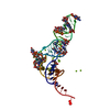
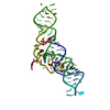
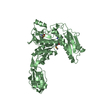
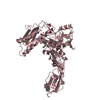
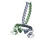
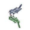
| Title | CRYSTALLOGRAPHIC AND BIOCHEMICAL INVESTIGATION OF THE LEAD(II)-CATALYZED HYDROLYSIS OF YEAST PHENYLALANINE TRNA | ||||||
 Components Components | TRNAPHE | ||||||
 Keywords Keywords | RNA / TRANSLATION | ||||||
| Function / homology | LEAD (II) ION / SPERMINE / RNA / RNA (> 10) Function and homology information Function and homology information | ||||||
| Biological species |  | ||||||
| Method |  X-RAY DIFFRACTION / Resolution: 3 Å X-RAY DIFFRACTION / Resolution: 3 Å | ||||||
 Authors Authors | Dewan, J.C. / Brown, R.S. / Hingerty, B.E. / Klug, A. | ||||||
 Citation Citation | Journal: Biochemistry / Year: 1985 Title: Crystallographic and biochemical investigation of the lead(II)-catalyzed hydrolysis of yeast phenylalanine tRNA. Authors: Brown, R.S. / Dewan, J.C. / Klug, A. #1: Journal: Nature / Year: 1983Title: Pb(II)-Catalysed Cleavage of the Sugar-Phosphate Backbone of Yeast tRNA-Phe-Implications for Lead Toxicity and Self-Splicing RNA Authors: Brown, R.S. / Hingerty, B.E. / Dewan, J.C. / Klug, A. | ||||||
| History |
|
- Structure visualization
| Structure viewer | Molecule: MolmilJmol/JSmol |
|---|
- Downloads & links
-Download
| PDBx/mmCIF format | 1tn1.cif.gz | 69 KB | Display | PDBx/mmCIF format |
|---|---|---|---|---|
| PDB format | pdb1tn1.ent.gz | 41.3 KB | Display | PDB format |
| PDBx/mmJSON format | 1tn1.json.gz | Tree view | PDBx/mmJSON format | |
| Others |  Other downloads Other downloads |
-Validation report
| Arichive directory | https://data.pdbj.org/pub/pdb/validation_reports/tn/1tn1ftp://data.pdbj.org/pub/pdb/validation_reports/tn/1tn1 | HTTPS FTP |
|---|
-Related structure data


-Links
 PDBj
PDBj


- Assembly
| Deposited unit | 
| ||||||||
|---|---|---|---|---|---|---|---|---|---|
| 1 |
| ||||||||
| Unit cell |
|
-Components
| #1: RNA chain | Mass: 24890.121 Da / Num. of mol.: 1 / Source method: isolated from a natural source / Source: (natural) | ||||||
|---|---|---|---|---|---|---|---|
| #2: Chemical | ChemComp-SPM /   Mass: 202.340 Da / Num. of mol.: 1 / Source method: obtained synthetically / Formula: C10H26N4 Mass: 202.340 Da / Num. of mol.: 1 / Source method: obtained synthetically / Formula: C10H26N4 | ||||||
| #3: Chemical | ChemComp-MG /   Mass: 24.305 Da / Num. of mol.: 5 / Source method: obtained synthetically / Formula: Mg Mass: 24.305 Da / Num. of mol.: 5 / Source method: obtained synthetically / Formula: Mg#4: Chemical |   Mass: 207.200 Da / Num. of mol.: 3 / Source method: obtained synthetically / Formula: Pb Mass: 207.200 Da / Num. of mol.: 3 / Source method: obtained synthetically / Formula: Pb#5: Water | ChemComp-HOH / |  Mass: 18.015 Da / Num. of mol.: 107 / Source method: isolated from a natural source / Formula: H2O Mass: 18.015 Da / Num. of mol.: 107 / Source method: isolated from a natural source / Formula: H2ONonpolymer details | PB(1) IS BOUND IN THE T-PSI-C LOOP. PB(2) IS BOUND IN THE EXTRA LOOP REGION. PB(3) IS BOUND IN THE ...PB(1) IS BOUND IN THE T-PSI-C LOOP. PB(2) IS BOUND IN THE EXTRA LOOP REGION. PB(3) IS BOUND IN THE ANTICODON LOOP. THE COORDINATI | |
-Experimental details
-Experiment
| Experiment | Method: X-RAY DIFFRACTION |
|---|
- Sample preparation
| Crystal | Density Matthews: 2.38 Å3/Da / Density % sol: 48.37 % |
|---|
-Data collection
| Radiation | Monochromatic (M) / Laue (L): M / Scattering type: x-ray |
|---|---|
| Radiation wavelength | Relative weight: 1 |
| Reflection | *PLUS Num. obs: 4821 / Num. measured all: 10704 |
- Processing
| Software | Name: EREF / Classification: refinement | ||||||||||||
|---|---|---|---|---|---|---|---|---|---|---|---|---|---|
| Refinement | Rfactor Rwork: 0.227 / Highest resolution: 3 Å Details: AT PRESENT THE COORDINATES FOR THE PH7.4 PB-TRNA ARE NOT AVAILABLE. THUS ONLY THE COORDINATES FOR THE PB IONS ARE INCLUDED IN THIS ENTRY. REFINEMENT OF THE PH5.0 AND PH7.4 STRUCTURES ...Details: AT PRESENT THE COORDINATES FOR THE PH7.4 PB-TRNA ARE NOT AVAILABLE. THUS ONLY THE COORDINATES FOR THE PB IONS ARE INCLUDED IN THIS ENTRY. REFINEMENT OF THE PH5.0 AND PH7.4 STRUCTURES INDICATED THAT THEY CHANGED VERY LITTLE FROM THE NATIVE STRUCTURE. THE ONLY REAL DIFFERENCE IS THE PRESENCE OF THE PB IONS. ALSO, IN THE PH 7.4 STRUCTURE, THE SUGAR-PHOSPHATE BACKBONE IS CLEAVED BETWEEN RESIDUES H2U 17 AND G 18. THIS ENTRY ORIGINALLY CONTAINED ONLY THE COORDINATES OF THE THREE PB IONS. IN ORDER TO BRING COMPLETENESS TO THE STRUCTURE, THE ENTRY HAS BEEN REMEDIATED BY MERGING THE COORDINATES OF THE TRNA, MG ION AND WATER FROM THE RELATED ENTRY 1TN2. | ||||||||||||
| Refinement step | Cycle: LAST / Highest resolution: 3 Å
| ||||||||||||
| Refinement | *PLUS Highest resolution: 3 Å | ||||||||||||
| Solvent computation | *PLUS | ||||||||||||
| Displacement parameters | *PLUS |
