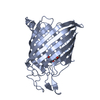
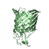
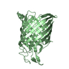
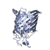
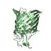
- PDB-1tlw: Tsx structure complexed with thymidine -
+
Open data
ID or keywords:
Loading...
-
Basic information
Entry
Database: PDB / ID: 1tlw
Title
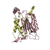
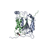
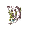
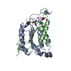
Tsx structure complexed with thymidine
Components
Nucleoside-specific channel-forming protein tsx
Keywords
MEMBRANE PROTEIN / nucleoside transporter / BETA BARREL / thymidine
Function / homology
Function and homology information
nucleoside-specific channel forming porin activity / nucleoside transport / pore complex / monoatomic ion transport / cell outer membrane / symbiont entry into host cell Similarity search - Function
Type: DNA OH 5 prime terminus / Mass: 242.229 Da / Num. of mol.: 4 / Source method: obtained synthetically / Formula: C10H14N2O5
-
Experimental details
-
Experiment
Experiment
Method: X-RAY DIFFRACTION / Number of used crystals: 1
-
Sample preparation
Crystal
Density Matthews: 6.2 Å3/Da / Density % sol: 82 %
Crystal grow
Temperature: 286 K / Method: soaking the crystal with 10 mm thymidine / pH: 4.3 Details: PEG550 27-32%, sodium acetate 50 mM, glycerol 10%, pH 4.3, soaking the crystal with 10 mM thymidine, temperature 286K
In the structure databanks used in Yorodumi, some data are registered as the other names, "COVID-19 virus" and "2019-nCoV". Here are the details of the virus and the list of structure data.
Jan 31, 2019. EMDB accession codes are about to change! (news from PDBe EMDB page)
EMDB accession codes are about to change! (news from PDBe EMDB page)
The allocation of 4 digits for EMDB accession codes will soon come to an end. Whilst these codes will remain in use, new EMDB accession codes will include an additional digit and will expand incrementally as the available range of codes is exhausted. The current 4-digit format prefixed with “EMD-” (i.e. EMD-XXXX) will advance to a 5-digit format (i.e. EMD-XXXXX), and so on. It is currently estimated that the 4-digit codes will be depleted around Spring 2019, at which point the 5-digit format will come into force.
The EM Navigator/Yorodumi systems omit the EMD- prefix.
Related info.:Q: What is EMD? / ID/Accession-code notation in Yorodumi/EM Navigator
Yorodumi is a browser for structure data from EMDB, PDB, SASBDB, etc.
This page is also the successor to EM Navigator detail page, and also detail information page/front-end page for Omokage search.
The word "yorodu" (or yorozu) is an old Japanese word meaning "ten thousand". "mi" (miru) is to see.
Related info.:EMDB / PDB / SASBDB / Comparison of 3 databanks / Yorodumi Search / Aug 31, 2016. New EM Navigator & Yorodumi / Yorodumi Papers / Jmol/JSmol / Function and homology information / Changes in new EM Navigator and Yorodumi
 Movie
Movie Controller


 Open data
 Basic information
 Components
 Keywords
 Function and homology information
 X-RAY DIFFRACTION /
  Authors
 Citation
 Structure visualization
 Downloads & links
 Other downloads
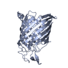


 PDBj
PDBj Assembly
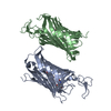

 Type: DNA OH 5 prime terminus / Mass: 242.229 Da / Num. of mol.: 4 / Source method: obtained synthetically / Formula: C10H14N2O5
 Sample preparation
 / Beamline: X25 / Wavelength: 0.979 Å
 Processing