[English] 日本語
Yorodumi
- PDB-1teg: Crystal structure of the spinach plastocyanin mutants G8D/K30C/T6... -

+
Open data


ID or keywords:

Loading...

-
Basic information

Entry
Database: PDB / ID: 1teg
Title
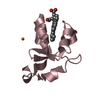
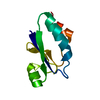
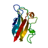
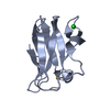
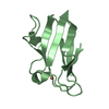
Crystal structure of the spinach plastocyanin mutants G8D/K30C/T69C and K30C/T69C- a study of the effect on crystal packing and thermostability from the introduction of a novel disulfide bond
ComponentsPlastocyanin, chloroplast
KeywordsPHOTOSYNTHESIS / Plastocyanin / disulfide bond / mutant / blue copper protein
Function / homology
Function and homology information


: / chloroplast thylakoid lumen / chloroplast thylakoid membrane / copper ion binding
Similarity search - Function
Plastocyanin / Blue (type 1) copper protein, plastocyanin-type / Blue (type 1) copper domain / Copper binding proteins, plastocyanin/azurin family / Blue (type 1) copper protein, binding site / Type-1 copper (blue) proteins signature. / Cupredoxins - blue copper proteins / Cupredoxin / Immunoglobulin-like / Sandwich / Mainly Beta
Similarity search - Domain/homology
COPPER (II) ION / Plastocyanin, chloroplastic
Similarity search - Component
Biological speciesSpinacia oleracea (spinach)
MethodX-RAY DIFFRACTION / SYNCHROTRON / ISOMORPHOUS STRUCTURE / Resolution: 1.96 Å
AuthorsOkvist, M. / Jacobson, F. / Jansson, H. / Hansson, O. / Sjolin, L.
CitationJournal: To be Published
Title: Novel Disulfide Bonds Effect the Thermostability of Plastocyanin. Crystal structures of the triple plastocyanin mutant G8D/K30C/T69C and the double plastocyanin mutant K30C/T69C from spinach ...Title: Novel Disulfide Bonds Effect the Thermostability of Plastocyanin. Crystal structures of the triple plastocyanin mutant G8D/K30C/T69C and the double plastocyanin mutant K30C/T69C from spinach at 1.90 A and 1.96 A resolution, respectively.
Authors: Okvist, M. / Jacobson, F. / Jansson, H. / Hansson, O. / Sjolin, L.
History
DepositionMay 25, 2004Deposition site: RCSB / Processing site: RCSB
Revision 1.0Nov 1, 2005Provider: repository / Type: Initial release
Revision 1.1Apr 30, 2008Group: Version format compliance
Revision 1.2Jul 13, 2011Group: Version format compliance
Revision 1.3Nov 16, 2011Group: Atomic model
Revision 1.4Mar 7, 2018Group: Data collection / Category: diffrn_source / Item: _diffrn_source.pdbx_synchrotron_site
Revision 1.5Oct 27, 2021Group: Database references / Derived calculations
Category: database_2 / struct_conn ...database_2 / struct_conn / struct_ref_seq_dif / struct_site
Item: _database_2.pdbx_DOI / _database_2.pdbx_database_accession ..._database_2.pdbx_DOI / _database_2.pdbx_database_accession / _struct_conn.ptnr1_auth_comp_id / _struct_conn.ptnr1_auth_seq_id / _struct_conn.ptnr1_label_asym_id / _struct_conn.ptnr1_label_atom_id / _struct_conn.ptnr1_label_comp_id / _struct_conn.ptnr1_label_seq_id / _struct_conn.ptnr2_auth_comp_id / _struct_conn.ptnr2_auth_seq_id / _struct_conn.ptnr2_label_asym_id / _struct_conn.ptnr2_label_atom_id / _struct_conn.ptnr2_label_comp_id / _struct_conn.ptnr2_label_seq_id / _struct_ref_seq_dif.details / _struct_site.pdbx_auth_asym_id / _struct_site.pdbx_auth_comp_id / _struct_site.pdbx_auth_seq_id
Revision 1.6Aug 23, 2023Group: Data collection / Refinement description
Category: chem_comp_atom / chem_comp_bond / pdbx_initial_refinement_model
Revision 1.7Nov 20, 2024Group: Structure summary / Category: pdbx_entry_details / pdbx_modification_feature

-
Structure visualization

Structure viewerMolecule:
MolmilJmol/JSmol

Downloads & links

-
Assembly

Deposited unit
A: Plastocyanin, chloroplast
B: Plastocyanin, chloroplast
hetero molecules


Theoretical massNumber of molelcules
Total (without water)20,9565
Polymers20,7932
Non-polymers1633
Water1,54986
1
A: Plastocyanin, chloroplast
hetero molecules


Theoretical massNumber of molelcules
Total (without water)10,4963
Polymers10,3971
Non-polymers992
Water181
TypeNameSymmetry operationNumber
identity operation1_555x,y,z1
2
B: Plastocyanin, chloroplast
hetero molecules


Theoretical massNumber of molelcules
Total (without water)10,4602
Polymers10,3971
Non-polymers641
Water181
TypeNameSymmetry operationNumber
identity operation1_555x,y,z1
Unit cell
Length a, b, c (Å)52.196, 52.196, 126.928
Angle α, β, γ (deg.)90.00, 90.00, 120.00
Int Tables number154
Space group name H-MP3221
Components on special symmetry positions
IDModelComponents
11B-138-

HOH

21B-141-

HOH

-
Components

#1: Protein Plastocyanin, chloroplast


Mass: 10396.559 Da / Num. of mol.: 2 / Mutation: K30C, T69C
Source method: isolated from a genetically manipulated source
Source: (gene. exp.) Spinacia oleracea (spinach) / Gene: PETE / Plasmid: pUG101tr / Production host: Escherichia coli (E. coli) / Strain (production host): RV308 / References: UniProt: P00289
#2: Chemical ChemComp-CU / COPPER (II) ION


Mass: 63.546 Da / Num. of mol.: 2 / Source method: obtained synthetically / Formula: Cu
#3: Chemical ChemComp-CL / CHLORIDE ION


Mass: 35.453 Da / Num. of mol.: 1 / Source method: obtained synthetically / Formula: Cl
#4: Water ChemComp-HOH / water


Mass: 18.015 Da / Num. of mol.: 86 / Source method: isolated from a natural source / Formula: H2O
Has protein modificationY

-
Experimental details

-
Experiment

ExperimentMethod: X-RAY DIFFRACTION / Number of used crystals: 1

-
Sample preparation

CrystalDensity Matthews: 2.4 Å3/Da / Density % sol: 48.73 %
Crystal growTemperature: 293 K / Method: vapor diffusion, hanging drop / pH: 4.5
Details: 21% PEG 3350, 0.25M MgCl2, 0.1M Na-acetate, pH 4.5, VAPOR DIFFUSION, HANGING DROP, temperature 293.0K

-
Data collection

DiffractionMean temperature: 100 K
Diffraction sourceSource: SYNCHROTRON / Site: MAX II / Beamline: I711 / Wavelength: 1.095 Å
DetectorType: MARRESEARCH / Detector: CCD / Date: Apr 25, 2002
RadiationMonochromator: FOCUSED Si(111) / Protocol: SINGLE WAVELENGTH / Monochromatic (M) / Laue (L): M / Scattering type: x-ray
Radiation wavelengthWavelength: 1.095 Å / Relative weight: 1
ReflectionResolution: 1.96→45.2 Å / Num. all: 14601 / Num. obs: 14601 / % possible obs: 98.1 % / Observed criterion σ(F): -99.9 / Observed criterion σ(I): -3.7 / Redundancy: 10.83 % / Biso Wilson estimate: 11.3 Å2 / Rmerge(I) obs: 0.05 / Net I/σ(I): 34.15
Reflection shellResolution: 1.96→2.07 Å / Redundancy: 6.86 % / Rmerge(I) obs: 0.1 / Mean I/σ(I) obs: 6.97 / % possible all: 90.69

-
Processing

Software
NameVersionClassification
CNS1.1refinement
MOSFLMdata reduction
CCP4(SCALA)data scaling
RefinementMethod to determine structure: ISOMORPHOUS STRUCTURE
Starting model: PDB entry 1tef - G8D,K30C,T69C tripple mutant.
Resolution: 1.96→45.2 Å / Rfactor Rfree error: 0.011 / Data cutoff high absF: 1349137.12 / Data cutoff low absF: 0 / Isotropic thermal model: RESTRAINED / Cross valid method: THROUGHOUT / σ(F): 0 / Stereochemistry target values: Engh & Huber / Details: BULK SOLVENT MODEL USED
RfactorNum. reflection% reflectionSelection details
Rfree0.304 707 4.8 %RANDOM
Rwork0.287 ---
obs0.287 14601 97.6 %-
all-14601 --
Solvent computationSolvent model: FLAT MODEL / Bsol: 40.6002 Å2 / ksol: 0.356242 e/Å3
Displacement parametersBiso mean: 19 Å2
Baniso -1Baniso -2Baniso -3
1-1.28 Å2-0.1 Å20 Å2
2--1.28 Å20 Å2
3----2.56 Å2
Refine analyze
FreeObs
Luzzati coordinate error0.36 Å0.32 Å
Luzzati d res low-5 Å
Luzzati sigma a0.24 Å0.18 Å
Refinement stepCycle: LAST / Resolution: 1.96→45.2 Å
ProteinNucleic acidLigandSolventTotal
Num. atoms1458 0 3 86 1547
Refine LS restraints
Refine-IDTypeDev idealDev ideal target
X-RAY DIFFRACTIONc_bond_d0.012
X-RAY DIFFRACTIONc_bond_d_na
X-RAY DIFFRACTIONc_bond_d_prot
X-RAY DIFFRACTIONc_angle_d
X-RAY DIFFRACTIONc_angle_d_na
X-RAY DIFFRACTIONc_angle_d_prot
X-RAY DIFFRACTIONc_angle_deg1.6
X-RAY DIFFRACTIONc_angle_deg_na
X-RAY DIFFRACTIONc_angle_deg_prot
X-RAY DIFFRACTIONc_dihedral_angle_d27.7
X-RAY DIFFRACTIONc_dihedral_angle_d_na
X-RAY DIFFRACTIONc_dihedral_angle_d_prot
X-RAY DIFFRACTIONc_improper_angle_d0.94
X-RAY DIFFRACTIONc_improper_angle_d_na
X-RAY DIFFRACTIONc_improper_angle_d_prot
X-RAY DIFFRACTIONc_mcbond_it0.831.5
X-RAY DIFFRACTIONc_mcangle_it1.332
X-RAY DIFFRACTIONc_scbond_it1.22
X-RAY DIFFRACTIONc_scangle_it1.82.5
LS refinement shellResolution: 1.96→2.07 Å / Rfactor Rfree error: 0.033 / Total num. of bins used: 6
RfactorNum. reflection% reflection
Rfree0.335 100 5.1 %
Rwork0.308 1846 -
obs--78.1 %
Xplor file
Refine-IDSerial noParam fileTopol file
X-RAY DIFFRACTION1PROTEIN_REP.PARAMPROTEIN.TOP
X-RAY DIFFRACTION2ION.PARAMWATER.TOP
X-RAY DIFFRACTION3WATER.PARAMION.TOP

+
About Yorodumi

-
News

-
Feb 9, 2022. New format data for meta-information of EMDB entries

New format data for meta-information of EMDB entries

  • Version 3 of the EMDB header file is now the official format.
  • The previous official version 1.9 will be removed from the archive.

Related info.:EMDB header

External links:wwPDB to switch to version 3 of the EMDB data model

-
Aug 12, 2020. Covid-19 info

Covid-19 info

URL: https://pdbj.org/emnavi/covid19.php

New page: Covid-19 featured information page in EM Navigator.

Related info.:Covid-19 info / Mar 5, 2020. Novel coronavirus structure data

+
Mar 5, 2020. Novel coronavirus structure data

Novel coronavirus structure data

Related info.:Yorodumi Speices / Aug 12, 2020. Covid-19 info

External links:COVID-19 featured content - PDBj / Molecule of the Month (242):Coronavirus Proteases

+
Jan 31, 2019. EMDB accession codes are about to change! (news from PDBe EMDB page)

EMDB accession codes are about to change! (news from PDBe EMDB page)

  • The allocation of 4 digits for EMDB accession codes will soon come to an end. Whilst these codes will remain in use, new EMDB accession codes will include an additional digit and will expand incrementally as the available range of codes is exhausted. The current 4-digit format prefixed with “EMD-” (i.e. EMD-XXXX) will advance to a 5-digit format (i.e. EMD-XXXXX), and so on. It is currently estimated that the 4-digit codes will be depleted around Spring 2019, at which point the 5-digit format will come into force.
  • The EM Navigator/Yorodumi systems omit the EMD- prefix.

Related info.:Q: What is EMD? / ID/Accession-code notation in Yorodumi/EM Navigator

External links:EMDB Accession Codes are Changing Soon! / Contact to PDBj

+
Jul 12, 2017. Major update of PDB

Major update of PDB

  • wwPDB released updated PDB data conforming to the new PDBx/mmCIF dictionary.
  • This is a major update changing the version number from 4 to 5, and with Remediation, in which all the entries are updated.
  • In this update, many items about electron microscopy experimental information are reorganized (e.g. em_software).
  • Now, EM Navigator and Yorodumi are based on the updated data.

External links:wwPDB Remediation / Enriched Model Files Conforming to OneDep Data Standards Now Available in the PDB FTP Archive

-
Yorodumi

Thousand views of thousand structures

  • Yorodumi is a browser for structure data from EMDB, PDB, SASBDB, etc.
  • This page is also the successor to EM Navigator detail page, and also detail information page/front-end page for Omokage search.
  • The word "yorodu" (or yorozu) is an old Japanese word meaning "ten thousand". "mi" (miru) is to see.

Related info.:EMDB / PDB / SASBDB / Comparison of 3 databanks / Yorodumi Search / Aug 31, 2016. New EM Navigator & Yorodumi / Yorodumi Papers / Jmol/JSmol / Function and homology information / Changes in new EM Navigator and Yorodumi

Read more