 Movie
Movie Controller

[English] 日本語
 Yorodumi
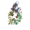
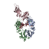
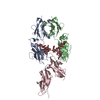
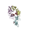
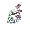
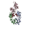
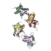
- PDB-1t89: CRYSTAL STRUCTURE OF A HUMAN TYPE III FC GAMMA RECEPTOR IN COMPLE... -
+ Open data

- Basic information
| Entry | Database: PDB / ID: 1t89 | ||||||||||||
|---|---|---|---|---|---|---|---|---|---|---|---|---|---|
| Title | CRYSTAL STRUCTURE OF A HUMAN TYPE III FC GAMMA RECEPTOR IN COMPLEX WITH AN FC FRAGMENT OF IGG1 (HEXAGONAL) | ||||||||||||
 Components Components |
| ||||||||||||
 Keywords Keywords | IMMUNE SYSTEM / Fc gamma receptor / IgG1 / CD16 / FcgRIII / immunoglobulin | ||||||||||||
| Function / homology |  Function and homology information Function and homology informationGPI anchor binding / IgG receptor activity / Post-translational modification: synthesis of GPI-anchored proteins / Fc-gamma receptor I complex binding / complement-dependent cytotoxicity / IgG immunoglobulin complex / antibody-dependent cellular cytotoxicity / IgG binding / immunoglobulin receptor binding / immunoglobulin complex, circulating ...GPI anchor binding / IgG receptor activity / Post-translational modification: synthesis of GPI-anchored proteins / Fc-gamma receptor I complex binding / complement-dependent cytotoxicity / IgG immunoglobulin complex / antibody-dependent cellular cytotoxicity / IgG binding / immunoglobulin receptor binding / immunoglobulin complex, circulating / Classical antibody-mediated complement activation / Initial triggering of complement / FCGR activation / complement activation, classical pathway / Role of phospholipids in phagocytosis / antigen binding / FCGR3A-mediated IL10 synthesis / secretory granule membrane / Regulation of Complement cascade / B cell receptor signaling pathway / FCGR3A-mediated phagocytosis / Regulation of actin dynamics for phagocytic cup formation / antibacterial humoral response / Interleukin-4 and Interleukin-13 signaling / blood microparticle / adaptive immune response / cell surface receptor signaling pathway / immune response / external side of plasma membrane / Neutrophil degranulation / extracellular space / extracellular exosome / extracellular region / plasma membrane Similarity search - Function | ||||||||||||
| Biological species |  Homo sapiens (human) Homo sapiens (human) | ||||||||||||
| Method |  X-RAY DIFFRACTION / SYNCHROTRON / MOLECULAR REPLACEMENT / Resolution: 3.5 Å X-RAY DIFFRACTION / SYNCHROTRON / MOLECULAR REPLACEMENT / Resolution: 3.5 Å | ||||||||||||
 Authors Authors | Radaev, S. / Motyka, S. / Fridman, W.-H. / Sautes-Fridman, C. / Sun, P.D. | ||||||||||||
 Citation Citation | Journal: J.Biol.Chem. / Year: 2001 Title: The structure of a human type III Fcgamma receptor in complex with Fc Authors: Radaev, S. / Motyka, S. / Fridman, W.-H. / Sautes-Fridman, C. / Sun, P.D. | ||||||||||||
| History |
|
- Structure visualization
| Structure viewer | Molecule: MolmilJmol/JSmol |
|---|
- Downloads & links
-Download
| PDBx/mmCIF format | 1t89.cif.gz | 139.5 KB | Display | PDBx/mmCIF format |
|---|---|---|---|---|
| PDB format | pdb1t89.ent.gz | 108.2 KB | Display | PDB format |
| PDBx/mmJSON format | 1t89.json.gz | Tree view | PDBx/mmJSON format | |
| Others |  Other downloads Other downloads |
-Validation report
| Arichive directory | https://data.pdbj.org/pub/pdb/validation_reports/t8/1t89ftp://data.pdbj.org/pub/pdb/validation_reports/t8/1t89 | HTTPS FTP |
|---|
-Related structure data
| Related structure data | 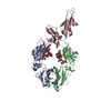 1t83C  1fc1S  1fnlS C: citing same article ( S: Starting model for refinement |
|---|---|
| Similar structure data |


-Links
 PDBj
PDBj


- Assembly
| Deposited unit | 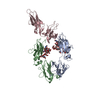
| ||||||||
|---|---|---|---|---|---|---|---|---|---|
| 1 |
| ||||||||
| Unit cell |
|
-Components
| #1: Antibody | Mass: 25267.645 Da / Num. of mol.: 2 / Fragment: Fc fragment of human IgG1 / Source method: isolated from a natural source / Source: (natural) Homo sapiens (human) / References: GenBank: 9857753, UniProt: P01857*PLUS#2: Protein | | Mass: 20103.416 Da / Num. of mol.: 1 / Fragment: Fc gamma receptor type III Source method: isolated from a genetically manipulated source Source: (gene. exp.) Homo sapiens (human) / Plasmid: pET-28b / Species (production host): Escherichia coli / Production host:  #3: Polysaccharide | beta-D-galactopyranose-(1-4)-2-acetamido-2-deoxy-alpha-D-glucopyranose-(1-2)-alpha-D-mannopyranose- ...beta-D-galactopyranose-(1-4)-2-acetamido-2-deoxy-alpha-D-glucopyranose-(1-2)-alpha-D-mannopyranose-(1-6)-[beta-D-mannopyranose-(1-3)]beta-D-mannopyranose-(1-4)-2-acetamido-2-deoxy-beta-D-glucopyranose-(1-4)-[beta-L-fucopyranose-(1-6)]2-acetamido-2-deoxy-beta-D-glucopyranose | Source method: isolated from a genetically manipulated source #4: Polysaccharide | 2-acetamido-2-deoxy-beta-D-glucopyranose-(1-2)-alpha-D-mannopyranose-(1-3)-[2-acetamido-2-deoxy- ...2-acetamido-2-deoxy-beta-D-glucopyranose-(1-2)-alpha-D-mannopyranose-(1-3)-[2-acetamido-2-deoxy-beta-D-glucopyranose-(1-2)-alpha-D-mannopyranose-(1-6)]beta-D-mannopyranose-(1-4)-2-acetamido-2-deoxy-alpha-D-glucopyranose-(1-4)-[alpha-L-fucopyranose-(1-6)]2-acetamido-2-deoxy-beta-D-glucopyranose | Source method: isolated from a genetically manipulated source Has protein modification | Y | |
|---|
-Experimental details
-Experiment
| Experiment | Method: X-RAY DIFFRACTION / Number of used crystals: 1 |
|---|
- Sample preparation
| Crystal | Density Matthews: 4 Å3/Da / Density % sol: 69 % |
|---|---|
| Crystal grow | Temperature: 298 K / Method: vapor diffusion / pH: 6 Details: PEG4000, HEPES, pH 6.0, VAPOR DIFFUSION, temperature 298.0K |
-Data collection
| Diffraction | Mean temperature: 100 K |
|---|---|
| Diffraction source | Source: SYNCHROTRON / Site: NSLS  / Beamline: X9B / Wavelength: 1 Å / Beamline: X9B / Wavelength: 1 Å |
| Detector | Type: ADSC QUANTUM 4 / Detector: CCD |
| Radiation | Protocol: SINGLE WAVELENGTH / Monochromatic (M) / Laue (L): M / Scattering type: x-ray |
| Radiation wavelength | Wavelength: 1 Å / Relative weight: 1 |
| Reflection | Resolution: 3.5→100 Å / Num. obs: 15541 / % possible obs: 98.7 % / Observed criterion σ(I): -3 / Redundancy: 4.7 % / Biso Wilson estimate: 82 Å2 / Rsym value: 0.087 / Net I/σ(I): 20.2 |
| Reflection shell | Resolution: 3.5→3.6 Å / Redundancy: 4.8 % / Mean I/σ(I) obs: 4 / Rsym value: 0.42 / % possible all: 99.4 |
- Processing
| Software |
| ||||||||||||||||||||
|---|---|---|---|---|---|---|---|---|---|---|---|---|---|---|---|---|---|---|---|---|---|
| Refinement | Method to determine structure: MOLECULAR REPLACEMENT Starting model: PDB ENTRY 1FNL AND 1FC1 Resolution: 3.5→10 Å / σ(F): 0 / Stereochemistry target values: Engh & Huber
| ||||||||||||||||||||
| Displacement parameters |
| ||||||||||||||||||||
| Refinement step | Cycle: LAST / Resolution: 3.5→10 Å
| ||||||||||||||||||||
| Refine LS restraints |
| ||||||||||||||||||||
| Xplor file |
|
