 Movie
Movie Controller

[English] 日本語
 Yorodumi
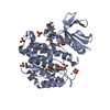
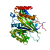
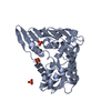
- PDB-1t6c: Structural characterization of the Ppx/GppA protein family: cryst... -
+ Open data

- Basic information
| Entry | Database: PDB / ID: 1t6c | ||||||
|---|---|---|---|---|---|---|---|
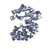
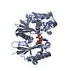
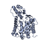
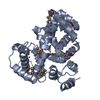
| Title | Structural characterization of the Ppx/GppA protein family: crystal structure of the Aquifex aeolicus family member | ||||||
 Components Components | exopolyphosphatase | ||||||
 Keywords Keywords | HYDROLASE / alpha/beta protein / Actin-like fold | ||||||
| Function / homology |  Function and homology information Function and homology information | ||||||
| Biological species |   Aquifex aeolicus (bacteria) Aquifex aeolicus (bacteria) | ||||||
| Method |  X-RAY DIFFRACTION / SYNCHROTRON / Molecular replacement with partial model, followed by the Arp/Warp procedure. / Resolution: 1.53 Å X-RAY DIFFRACTION / SYNCHROTRON / Molecular replacement with partial model, followed by the Arp/Warp procedure. / Resolution: 1.53 Å | ||||||
 Authors Authors | Kristensen, O. / Laurberg, M. / Liljas, A. / Kastrup, J.S. / Gajhede, M. | ||||||
 Citation Citation | Journal: Biochemistry / Year: 2004 Title: Structural characterization of the stringent response related exopolyphosphatase/guanosine pentaphosphate phosphohydrolase protein family Authors: Kristensen, O. / Laurberg, M. / Liljas, A. / Kastrup, J.S. / Gajhede, M. #1: Journal: Science / Year: 2001 Title: Role of inorganic polyphosphate in promoting ribosomal protein degradation by the Lon protease in E. coli Authors: Kuroda, A. / Nomura, K. / Ohtomo, R. / Kato, J. / Ikeda, T. / Takiguchi, N. / Ohtake, H. / Kornberg, A. #2: Journal: Proc.Natl.Acad.Sci.USA / Year: 1993 Title: Guanosine pentaphosphate phosphohydrolase of Escherichia coli is a long-chain exopolyphosphatase Authors: Keasling, J.D. / Bertsch, L. / Kornberg, A. #3: Journal: Trends Biochem.Sci. / Year: 1993 Title: Exopolyphosphate phosphatase and guanosine pentaphosphate phosphatase belong to the sugar kinase/actin/hsp 70 superfamily Authors: Reizer, J. / Reizer, A. / Saier Jr., M.H. / Bork, P. / Sander, C. | ||||||
| History |
|
- Structure visualization
| Structure viewer | Molecule: MolmilJmol/JSmol |
|---|
- Downloads & links
-Download
| PDBx/mmCIF format | 1t6c.cif.gz | 87.5 KB | Display | PDBx/mmCIF format |
|---|---|---|---|---|
| PDB format | pdb1t6c.ent.gz | 66 KB | Display | PDB format |
| PDBx/mmJSON format | 1t6c.json.gz | Tree view | PDBx/mmJSON format | |
| Others |  Other downloads Other downloads |
-Validation report
| Arichive directory | https://data.pdbj.org/pub/pdb/validation_reports/t6/1t6cftp://data.pdbj.org/pub/pdb/validation_reports/t6/1t6c | HTTPS FTP |
|---|
-Related structure data


-Links
 PDBj
PDBj- Assembly
| Deposited unit | 
| ||||||||
|---|---|---|---|---|---|---|---|---|---|
| 1 |
| ||||||||
| Unit cell |
| ||||||||
| Details | The biological relevant unit is monomeric. |
-Components
-Protein , 1 types, 1 molecules A
| #1: Protein | Mass: 35831.332 Da / Num. of mol.: 1 Source method: isolated from a genetically manipulated source Source: (gene. exp.) Aquifex aeolicus (bacteria) / Strain: VF5 / Gene: ppx / Plasmid: pET28a / Production host: References: UniProt: O67040, exopolyphosphatase, guanosine-5'-triphosphate,3'-diphosphate phosphatase |
|---|
-Non-polymers , 5 types, 401 molecules 


| #2: Chemical | ChemComp-IOD /  Mass: 126.904 Da / Num. of mol.: 1 / Source method: obtained synthetically / Formula: I Mass: 126.904 Da / Num. of mol.: 1 / Source method: obtained synthetically / Formula: I | ||||
|---|---|---|---|---|---|
| #3: Chemical | ChemComp-CA /  Mass: 40.078 Da / Num. of mol.: 1 / Source method: obtained synthetically / Formula: Ca Mass: 40.078 Da / Num. of mol.: 1 / Source method: obtained synthetically / Formula: Ca | ||||
| #4: Chemical |  Mass: 35.453 Da / Num. of mol.: 3 / Source method: obtained synthetically / Formula: Cl Mass: 35.453 Da / Num. of mol.: 3 / Source method: obtained synthetically / Formula: Cl#5: Chemical | ChemComp-MPD / (  Mass: 118.174 Da / Num. of mol.: 6 / Source method: obtained synthetically / Formula: C6H14O2 / Comment: precipitant*YM Mass: 118.174 Da / Num. of mol.: 6 / Source method: obtained synthetically / Formula: C6H14O2 / Comment: precipitant*YM#6: Water | ChemComp-HOH / | Mass: 18.015 Da / Num. of mol.: 390 / Source method: isolated from a natural source / Formula: H2O |
-Experimental details
-Experiment
| Experiment | Method: X-RAY DIFFRACTION / Number of used crystals: 1 |
|---|
- Sample preparation
| Crystal | Density Matthews: 2.3 Å3/Da / Density % sol: 46.4 % |
|---|---|
| Crystal grow | Temperature: 293 K / Method: vapor diffusion, hanging drop / pH: 6.4 Details: MPD, MES, TRIS, KCl, DTT, The crystal was soaked in K2HgI4/KI containing solution, pH 6.4, VAPOR DIFFUSION, HANGING DROP, temperature 293K |
-Data collection
| Diffraction | Mean temperature: 100 K |
|---|---|
| Diffraction source | Source: SYNCHROTRON / Site: MAX II  / Beamline: I711 / Wavelength: 0.993 Å / Beamline: I711 / Wavelength: 0.993 Å |
| Detector | Type: MARRESEARCH / Detector: IMAGE PLATE / Date: Aug 13, 2000 |
| Radiation | Monochromator: BENDABLE ASYMMETRICALLY CUT SI(111) CRYSTAL IN COMBINATION WITH VERTICALLY FOCUSING MIRROR Protocol: SINGLE WAVELENGTH / Monochromatic (M) / Laue (L): M / Scattering type: x-ray |
| Radiation wavelength | Wavelength: 0.993 Å / Relative weight: 1 |
| Reflection | Resolution: 1.53→15 Å / Num. all: 47505 / Num. obs: 47505 / % possible obs: 95.8 % / Observed criterion σ(F): 0 / Observed criterion σ(I): 0 / Redundancy: 6.9 % / Biso Wilson estimate: 19 Å2 / Rmerge(I) obs: 0.069 / Rsym value: 0.061 / Net I/σ(I): 22.8 |
| Reflection shell | Resolution: 1.53→1.61 Å / Redundancy: 2.7 % / Rmerge(I) obs: 0.341 / Mean I/σ(I) obs: 2.2 / Num. unique all: 5936 / Rsym value: 0.24 / % possible all: 84.6 |
- Processing
| Software |
| ||||||||||||||||||||||||||||||||||||||||||||||||||||||||||||||||||||||||||||||||||||||||||||||||||||
|---|---|---|---|---|---|---|---|---|---|---|---|---|---|---|---|---|---|---|---|---|---|---|---|---|---|---|---|---|---|---|---|---|---|---|---|---|---|---|---|---|---|---|---|---|---|---|---|---|---|---|---|---|---|---|---|---|---|---|---|---|---|---|---|---|---|---|---|---|---|---|---|---|---|---|---|---|---|---|---|---|---|---|---|---|---|---|---|---|---|---|---|---|---|---|---|---|---|---|---|---|---|
| Refinement | Method to determine structure: Molecular replacement with partial model, followed by the Arp/Warp procedure. Starting model: A very incomplete model was obtained through experimental phasing and Arp/Warp tracing of the Type II crystal form Resolution: 1.53→15 Å / Cor.coef. Fo:Fc: 0.969 / Cor.coef. Fo:Fc free: 0.934 / SU B: 1.703 / SU ML: 0.058 / Isotropic thermal model: ISOTROPIC / Cross valid method: THROUGHOUT / σ(F): 0 / σ(I): 0 / ESU R: 0.073 / ESU R Free: 0.085 / Stereochemistry target values: MAXIMUM LIKELIHOOD
| ||||||||||||||||||||||||||||||||||||||||||||||||||||||||||||||||||||||||||||||||||||||||||||||||||||
| Solvent computation | Ion probe radii: 0.8 Å / Shrinkage radii: 0.8 Å / VDW probe radii: 1.4 Å / Solvent model: BABINET MODEL WITH MASK | ||||||||||||||||||||||||||||||||||||||||||||||||||||||||||||||||||||||||||||||||||||||||||||||||||||
| Displacement parameters | Biso mean: 16.249 Å2
| ||||||||||||||||||||||||||||||||||||||||||||||||||||||||||||||||||||||||||||||||||||||||||||||||||||
| Refinement step | Cycle: LAST / Resolution: 1.53→15 Å
| ||||||||||||||||||||||||||||||||||||||||||||||||||||||||||||||||||||||||||||||||||||||||||||||||||||
| Refine LS restraints |
| ||||||||||||||||||||||||||||||||||||||||||||||||||||||||||||||||||||||||||||||||||||||||||||||||||||
| LS refinement shell | Resolution: 1.53→1.57 Å / Total num. of bins used: 20
|
