 Movie
Movie Controller

[English] 日本語
 Yorodumi
- PDB-1t5r: STRUCTURE OF THE PANTON-VALENTINE LEUCOCIDIN S COMPONENT FROM STA... -
+ Open data

- Basic information
| Entry | Database: PDB / ID: 1t5r | ||||||
|---|---|---|---|---|---|---|---|
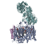
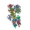
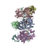
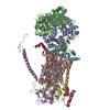
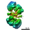
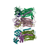
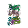
| Title | STRUCTURE OF THE PANTON-VALENTINE LEUCOCIDIN S COMPONENT FROM STAPHYLOCOCCUS AUREUS | ||||||
 Components Components | LukS-PV | ||||||
 Keywords Keywords | UNKNOWN FUNCTION / BI-COMPONENT LEUCOTOXIN / PORE-FORMING TOXIN / STAPHYLOCOCCUS AUREUS / S COMPONENT LEUCOCIDIN | ||||||
| Function / homology |  Function and homology information Function and homology information | ||||||
| Biological species |  Staphylococcus phage PVL (virus) Staphylococcus phage PVL (virus) | ||||||
| Method |  X-RAY DIFFRACTION / SYNCHROTRON / MOLECULAR REPLACEMENT / Resolution: 2 Å X-RAY DIFFRACTION / SYNCHROTRON / MOLECULAR REPLACEMENT / Resolution: 2 Å | ||||||
 Authors Authors | Guillet, V. / Roblin, P. / Keller, D. / Prevost, G. / Mourey, L. | ||||||
 Citation Citation | Journal: J.Biol.Chem. / Year: 2004 Title: Crystal structure of leucotoxin S component: new insight into the Staphylococcal beta-barrel pore-forming toxins. Authors: Guillet, V. / Roblin, P. / Werner, S. / Coraiola, M. / Menestrina, G. / Monteil, H. / Mourey, L. #1: Journal: Acta Crystallogr.,Sect.D / Year: 2004Title: CRYSTALLIZATION AND PRELIMINARY CRYSTALLOGRAPHIC DATA OF A LEUCOTOXIN S COMPONENT FROM STAPHYLOCOCCUS AUREUS Authors: GUILLET, V. / KELLER, D. / PREVOST, G. / MOUREY, L. #2: Journal: Structure / Year: 1999Title: THE STRUCTURE OF A STAPHYLOCOCCUS AUREUS LEUCOCIDIN COMPONENT (LUKF-PV) REVEALS THE FOLD OF THE WATER-SOLUBLE SPECIES OF A FAMILY OF TRANSMEMBRANE PORE-FORMING TOXINS. Authors: PEDELACQ, J.D. / MAVEYRAUD, L. / PREVOST, G. / BABA MOUSSA, L. / GONZALES, A. / COURCELLE, E. / SHEPARD, W. / MONTEIL, H. / SAMAMA, J.P. / MOUREY, L. | ||||||
| History |
|
- Structure visualization
| Structure viewer | Molecule: MolmilJmol/JSmol |
|---|
- Downloads & links
-Download
| PDBx/mmCIF format | 1t5r.cif.gz | 437.5 KB | Display | PDBx/mmCIF format |
|---|---|---|---|---|
| PDB format | pdb1t5r.ent.gz | 358.9 KB | Display | PDB format |
| PDBx/mmJSON format | 1t5r.json.gz | Tree view | PDBx/mmJSON format | |
| Others |  Other downloads Other downloads |
-Validation report
| Arichive directory | https://data.pdbj.org/pub/pdb/validation_reports/t5/1t5rftp://data.pdbj.org/pub/pdb/validation_reports/t5/1t5r | HTTPS FTP |
|---|
-Related structure data
| Related structure data | 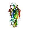 1pvlS S: Starting model for refinement |
|---|---|
| Similar structure data |

-Links
 PDBj
PDBj
- Assembly
| Deposited unit | 
| ||||||||
|---|---|---|---|---|---|---|---|---|---|
| 1 |
| ||||||||
| Unit cell |
|
-Components
| #1: Protein | Mass: 32355.621 Da / Num. of mol.: 8 / Source method: isolated from a natural source / Source: (natural) Staphylococcus phage PVL (virus) / Cellular location: CHROMOSOME / Strain: v8 / References: UniProt: O80066, UniProt: Q783R1*PLUS#2: Water | ChemComp-HOH / |  Mass: 18.015 Da / Num. of mol.: 821 / Source method: isolated from a natural source / Formula: H2O Mass: 18.015 Da / Num. of mol.: 821 / Source method: isolated from a natural source / Formula: H2O |
|---|
-Experimental details
-Experiment
| Experiment | Method: X-RAY DIFFRACTION / Number of used crystals: 1 |
|---|
- Sample preparation
| Crystal | Density Matthews: 2.7 Å3/Da / Density % sol: 54 % |
|---|---|
| Crystal grow | Temperature: 285 K / Method: vapor diffusion, hanging drop / pH: 8 Details: CRYSTALLIZATION CONDITIONS: VAPOR EQUILIBRIUM USING THE HANGING METHOD AT 285K. THE PROTEINS WERE CONCENTRATED AT 20-25 MG/ML IN MES-NAOH PH 6.0 (50MM), NACL (50MM). DROPS WERE PREPARED BY ...Details: CRYSTALLIZATION CONDITIONS: VAPOR EQUILIBRIUM USING THE HANGING METHOD AT 285K. THE PROTEINS WERE CONCENTRATED AT 20-25 MG/ML IN MES-NAOH PH 6.0 (50MM), NACL (50MM). DROPS WERE PREPARED BY MIXING EQUAL VOLUMES OF PROTEIN AND RESERVOIR SOLUTIONS AND USING RESERVOIR VOLUMES OF 500 microL CONTAINING 30% JEFFAMINE M-600, 0.1 M TRIS-HCL AT PH VALUES RANGING FROM 8.0 TO 8.9. MICRO- AND MACRO-SEEDING TECHNIQUES, INCLUDING CROSS-SEEDINGS, WERE USED TO GROW CRYSTALS. CRYSTAL SIZE (200X200X200 MM3) IN 6-10microL HANGING DROPS. , VAPOR DIFFUSION, HANGING DROP |
-Data collection
| Diffraction | Mean temperature: 100 K |
|---|---|
| Diffraction source | Source: SYNCHROTRON / Site: ESRF  / Beamline: ID14-1 / Wavelength: 0.934 Å / Beamline: ID14-1 / Wavelength: 0.934 Å |
| Detector | Type: ADSC QUANTUM 4 / Detector: CCD / Date: Nov 8, 2002 / Details: bent multilayer |
| Radiation | Monochromator: double crystal monochromator (thin diamond and focusing Germanium crystals) Protocol: SINGLE WAVELENGTH / Monochromatic (M) / Laue (L): M / Scattering type: x-ray |
| Radiation wavelength | Wavelength: 0.934 Å / Relative weight: 1 |
| Reflection | Resolution: 2→35.5 Å / Num. all: 169450 / Num. obs: 169450 / % possible obs: 93.6 % / Redundancy: 5.9 % / Biso Wilson estimate: 34.3 Å2 / Rsym value: 0.055 / Net I/σ(I): 9.1 |
| Reflection shell | Resolution: 2→2.11 Å / Redundancy: 2.5 % / Mean I/σ(I) obs: 1.6 / Num. unique all: 16649 / Rsym value: 0.47 / % possible all: 67.5 |
- Processing
| Software |
| ||||||||||||||||||||||||||||||||||||
|---|---|---|---|---|---|---|---|---|---|---|---|---|---|---|---|---|---|---|---|---|---|---|---|---|---|---|---|---|---|---|---|---|---|---|---|---|---|
| Refinement | Method to determine structure: MOLECULAR REPLACEMENT Starting model: PDB ENTRY 1PVL Resolution: 2→35.5 Å / Isotropic thermal model: anisotropic / Cross valid method: THROUGHOUT / σ(F): 3 / Stereochemistry target values: Engh & Huber
| ||||||||||||||||||||||||||||||||||||
| Displacement parameters | Biso mean: 36.3 Å2
| ||||||||||||||||||||||||||||||||||||
| Refine analyze |
| ||||||||||||||||||||||||||||||||||||
| Refinement step | Cycle: LAST / Resolution: 2→35.5 Å
| ||||||||||||||||||||||||||||||||||||
| Refine LS restraints |
| ||||||||||||||||||||||||||||||||||||
| LS refinement shell | Resolution: 2→2.13 Å
|
