[English] 日本語
Yorodumi
- PDB-1sy7: Crystal structure of the catalase-1 from Neurospora crassa, nativ... -

+
Open data


ID or keywords:

Loading...

-
Basic information

Entry
Database: PDB / ID: 1sy7
TitleCrystal structure of the catalase-1 from Neurospora crassa, native structure at 1.75A resolution.
ComponentsCatalase 1
KeywordsOXIDOREDUCTASE / Catalase / heme oxidation / singlet oxygen / Neurospora crassa
Function / homology
Function and homology information


conidium formation / ascospore wall / catalase / catalase activity / hydrogen peroxide catabolic process / response to oxidative stress / heme binding / extracellular region / metal ion binding / cytosol
Similarity search - Function
: / Catalase, four-helical domain / Catalase, mono-functional, haem-containing, clade 2 / Catalase, mono-functional, haem-containing, clade 2, helical domain / Hemocyanin, N-terminal domain / Catalase HpII, Chain A, domain 1 / Catalase core domain / Catalase haem-binding site / Catalase proximal heme-ligand signature. / Catalase ...: / Catalase, four-helical domain / Catalase, mono-functional, haem-containing, clade 2 / Catalase, mono-functional, haem-containing, clade 2, helical domain / Hemocyanin, N-terminal domain / Catalase HpII, Chain A, domain 1 / Catalase core domain / Catalase haem-binding site / Catalase proximal heme-ligand signature. / Catalase / Catalase active site / Catalase proximal active site signature. / Catalase immune-responsive domain / Catalase-related immune-responsive / Catalase core domain / Catalase, mono-functional, haem-containing / Catalase / catalase family profile. / DJ-1/PfpI / Catalase superfamily / DJ-1/PfpI family / Class I glutamine amidotransferase (GATase) domain / Class I glutamine amidotransferase-like / Up-down Bundle / Beta Barrel / Rossmann fold / 3-Layer(aba) Sandwich / Mainly Beta / Mainly Alpha / Alpha Beta
Similarity search - Domain/homology
CIS-HEME D HYDROXYCHLORIN GAMMA-SPIROLACTONE / PROTOPORPHYRIN IX CONTAINING FE / Catalase-1
Similarity search - Component
Biological speciesNeurospora crassa (fungus)
MethodX-RAY DIFFRACTION / SYNCHROTRON / MOLECULAR REPLACEMENT / Resolution: 1.75 Å
AuthorsDiaz, A. / Horjales, E. / Rudino-Pinera, E. / Arreola, R. / Hansberg, W.
CitationJournal: J.Mol.Biol. / Year: 2004
Title: Unusual Cys-Tyr covalent bond in a large catalase
Authors: Diaz, A. / Horjales, E. / Rudino-Pinera, E. / Arreola, R. / Hansberg, W.
History
DepositionApr 1, 2004Deposition site: RCSB / Processing site: RCSB
Revision 1.0Oct 12, 2004Provider: repository / Type: Initial release
Revision 1.1Apr 30, 2008Group: Version format compliance
Revision 1.2Jul 13, 2011Group: Derived calculations / Version format compliance
Revision 1.3Oct 11, 2017Group: Refinement description / Category: software / Item: _software.classification / _software.name
Revision 1.4Aug 23, 2023Group: Data collection / Database references ...Data collection / Database references / Derived calculations / Refinement description
Category: chem_comp_atom / chem_comp_bond ...chem_comp_atom / chem_comp_bond / database_2 / pdbx_initial_refinement_model / struct_conn / struct_site
Item: _database_2.pdbx_DOI / _database_2.pdbx_database_accession ..._database_2.pdbx_DOI / _database_2.pdbx_database_accession / _struct_conn.pdbx_leaving_atom_flag / _struct_site.pdbx_auth_asym_id / _struct_site.pdbx_auth_comp_id / _struct_site.pdbx_auth_seq_id
Remark 295 NON-CRYSTALLOGRAPHIC SYMMETRY THE TRANSFORMATIONS PRESENTED ON THE MTRIX RECORDS BELOW DESCRIBE ... NON-CRYSTALLOGRAPHIC SYMMETRY THE TRANSFORMATIONS PRESENTED ON THE MTRIX RECORDS BELOW DESCRIBE NON-CRYSTALLOGRAPHIC RELATIONSHIPS AMONG ATOMS IN THIS ENTRY. APPLYING THE APPROPRIATE MTRIX TRANSFORMATION TO THE RESIDUES LISTED FIRST WILL YIELD APPROXIMATE COORDINATES FOR THE RESIDUES LISTED SECOND. CHAIN IDENTIFIERS GIVEN AS "?" REFER TO CHAINS FOR WHICH ATOMS ARE NOT FOUND IN THIS ENTRY. APPLIED TO TRANSFORMED TO TRANSFORM CHAIN RESIDUES CHAIN RESIDUES RMSD 1 A 39 .. 716 B 39 .. 716 0.022
Remark 600
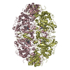
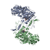
HETEROGEN THIS STRUCTURE PRESENTS AN ALTERNATE CONFORMATION OF THE HEME GROUP BETWEEN HEME B (HEM, ...HETEROGEN THIS STRUCTURE PRESENTS AN ALTERNATE CONFORMATION OF THE HEME GROUP BETWEEN HEME B (HEM, ALTERNATE CONFORMER A) AND HEME D (HDD, ALTERNATE CONFORMER B).

-
Structure visualization

Structure viewerMolecule:
MolmilJmol/JSmol

Downloads & links

-
Assembly

Deposited unit
A: Catalase 1
B: Catalase 1
hetero molecules


Theoretical massNumber of molelcules
Total (without water)163,0176
Polymers160,5192
Non-polymers2,4984
Water31,0761725
1
A: Catalase 1
B: Catalase 1
hetero molecules

A: Catalase 1
B: Catalase 1
hetero molecules


Theoretical massNumber of molelcules
Total (without water)326,03412
Polymers321,0384
Non-polymers4,9968
Water724
TypeNameSymmetry operationNumber
identity operation1_555x,y,z1
crystal symmetry operation2_555-x,y,-z1
Buried area55560 Å2
ΔGint-319 kcal/mol
Surface area77320 Å2
MethodPISA, PQS
Unit cell
Length a, b, c (Å)130.007, 182.242, 90.364
Angle α, β, γ (deg.)90.00, 133.41, 90.00
Int Tables number5
Space group name H-MC121
Noncrystallographic symmetry (NCS)NCS oper: (Code: given
Matrix: (-0.99523151, -0.00087905, 0.09753705), (0.00089592, -0.99999958, 0.00012913), (0.0975369, 0.00021589, 0.99523187)
Vector: -0.04701458, -0.79932737, 0.01917373)
Details
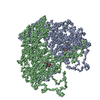
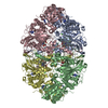
The biological unit is a homo-tetramer generated from the dimer in the asymmetric unit. To generate the tetramer (biological unit) apply the following matrix to chains A and B: -1.000000 0.000000 0.000000 0.00000 0.000000 1.000000 0.000000 0.00000 0.000000 0.000000 -1.000000 0.00000

-
Components

#1: Protein Catalase 1


Mass: 80259.438 Da / Num. of mol.: 2 / Source method: isolated from a natural source / Source: (natural) Neurospora crassa (fungus) / Strain: 74-ORS23-1A / References: UniProt: Q9C168, catalase
#2: Chemical ChemComp-HDD / CIS-HEME D HYDROXYCHLORIN GAMMA-SPIROLACTONE / HEME


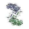
Mass: 632.487 Da / Num. of mol.: 2 / Source method: obtained synthetically / Formula: C34H32FeN4O5
#3: Chemical ChemComp-HEM / PROTOPORPHYRIN IX CONTAINING FE / HEME


Mass: 616.487 Da / Num. of mol.: 2 / Source method: obtained synthetically / Formula: C34H32FeN4O4
#4: Water ChemComp-HOH / water


Mass: 18.015 Da / Num. of mol.: 1725 / Source method: isolated from a natural source / Formula: H2O

-
Experimental details

-
Experiment

ExperimentMethod: X-RAY DIFFRACTION / Number of used crystals: 1

-
Sample preparation

CrystalDensity Matthews: 2.42 Å3/Da / Density % sol: 42.5 %
Crystal growTemperature: 291 K / Method: vapor diffusion, hanging drop / pH: 7.8
Details: Ammonium sulfate, pH 7.8, VAPOR DIFFUSION, HANGING DROP, temperature 291K

-
Data collection

DiffractionMean temperature: 100 K
Diffraction sourceSource: SYNCHROTRON / Site: SSRL / Beamline: BL9-1 / Wavelength: 0.782 Å
DetectorType: MARRESEARCH / Detector: IMAGE PLATE / Date: Mar 20, 1999
RadiationMonochromator: Mirrors / Protocol: SINGLE WAVELENGTH / Monochromatic (M) / Laue (L): M / Scattering type: x-ray
Radiation wavelengthWavelength: 0.782 Å / Relative weight: 1
ReflectionResolution: 1.75→51.09 Å / Num. all: 153020 / Num. obs: 153020 / % possible obs: 89 % / Observed criterion σ(F): 1 / Redundancy: 2.4 % / Biso Wilson estimate: 17.5 Å2 / Rsym value: 0.059 / Net I/σ(I): 8.6
Reflection shellResolution: 1.75→1.86 Å / Redundancy: 2.3 % / Mean I/σ(I) obs: 2.3 / Num. unique all: 20575 / Rsym value: 0.326 / % possible all: 89.8

-
Processing

Software
NameVersionClassification
CNS1refinement
MAR345data collection
SCALEPACKdata scaling
CNSphasing
RefinementMethod to determine structure: MOLECULAR REPLACEMENT
Starting model: 1GGE
Resolution: 1.75→15 Å / Rfactor Rfree error: 0.002 / Data cutoff high absF: 2990881.03 / Data cutoff low absF: 0 / Isotropic thermal model: RESTRAINED / Cross valid method: THROUGHOUT / σ(F): 0
Details: THE SULFUR ATOM OF THE RESIDUE CYS 356 FORMS AN UNUSUAL BOND WITH THE C BETA ATOM FROM RESIDUE TYR 379
RfactorNum. reflection% reflectionSelection details
Rfree0.206 13616 10 %RANDOM
Rwork0.183 ---
obs0.183 136420 89 %-
all-136420 --
Solvent computationSolvent model: FLAT MODEL / Bsol: 66.1702 Å2 / ksol: 0.377236 e/Å3
Displacement parametersBiso mean: 21.6 Å2
Baniso -1Baniso -2Baniso -3
1-6.44 Å20 Å2-1.45 Å2
2---3.43 Å20 Å2
3----3.01 Å2
Refine analyze
FreeObs
Luzzati coordinate error0.21 Å0.19 Å
Luzzati d res low-5 Å
Luzzati sigma a0.25 Å0.24 Å
Refinement stepCycle: LAST / Resolution: 1.75→15 Å
ProteinNucleic acidLigandSolventTotal
Num. atoms11050 0 174 1725 12949
Refine LS restraints
Refine-IDTypeDev idealDev ideal target
X-RAY DIFFRACTIONc_bond_d0.007
X-RAY DIFFRACTIONc_angle_deg1.4
X-RAY DIFFRACTIONc_dihedral_angle_d23
X-RAY DIFFRACTIONc_improper_angle_d1.19
X-RAY DIFFRACTIONc_mcbond_it0.981.5
X-RAY DIFFRACTIONc_mcangle_it1.412
X-RAY DIFFRACTIONc_scbond_it1.842
X-RAY DIFFRACTIONc_scangle_it2.62.5
LS refinement shellResolution: 1.75→1.86 Å / Rfactor Rfree error: 0.006 / Total num. of bins used: 6
RfactorNum. reflection% reflection
Rfree0.287 2244 9.8 %
Rwork0.274 20575 -
obs-136420 89.8 %
Xplor file
Refine-IDSerial noParam fileTopol file
X-RAY DIFFRACTION1PROTEIN_REP.PARAMPROTEIN.TOP
X-RAY DIFFRACTION2WATER_REP.PARAMWATER_REP.TOP
X-RAY DIFFRACTION3HEMO-D.PARAMHEMO-D.TOP
X-RAY DIFFRACTION4HEMO-B.PARAMHEMO-B.TOP

+
About Yorodumi

-
News

-
Feb 9, 2022. New format data for meta-information of EMDB entries

New format data for meta-information of EMDB entries

  • Version 3 of the EMDB header file is now the official format.
  • The previous official version 1.9 will be removed from the archive.

Related info.:EMDB header

External links:wwPDB to switch to version 3 of the EMDB data model

-
Aug 12, 2020. Covid-19 info

Covid-19 info

URL: https://pdbjlvh1.pdbj.org/emnavi/covid19.php

New page: Covid-19 featured information page in EM Navigator.

Related info.:Covid-19 info / Mar 5, 2020. Novel coronavirus structure data

+
Mar 5, 2020. Novel coronavirus structure data

Novel coronavirus structure data

Related info.:Yorodumi Speices / Aug 12, 2020. Covid-19 info

External links:COVID-19 featured content - PDBj / Molecule of the Month (242):Coronavirus Proteases

+
Jan 31, 2019. EMDB accession codes are about to change! (news from PDBe EMDB page)

EMDB accession codes are about to change! (news from PDBe EMDB page)

  • The allocation of 4 digits for EMDB accession codes will soon come to an end. Whilst these codes will remain in use, new EMDB accession codes will include an additional digit and will expand incrementally as the available range of codes is exhausted. The current 4-digit format prefixed with “EMD-” (i.e. EMD-XXXX) will advance to a 5-digit format (i.e. EMD-XXXXX), and so on. It is currently estimated that the 4-digit codes will be depleted around Spring 2019, at which point the 5-digit format will come into force.
  • The EM Navigator/Yorodumi systems omit the EMD- prefix.

Related info.:Q: What is EMD? / ID/Accession-code notation in Yorodumi/EM Navigator

External links:EMDB Accession Codes are Changing Soon! / Contact to PDBj

+
Jul 12, 2017. Major update of PDB

Major update of PDB

  • wwPDB released updated PDB data conforming to the new PDBx/mmCIF dictionary.
  • This is a major update changing the version number from 4 to 5, and with Remediation, in which all the entries are updated.
  • In this update, many items about electron microscopy experimental information are reorganized (e.g. em_software).
  • Now, EM Navigator and Yorodumi are based on the updated data.

External links:wwPDB Remediation / Enriched Model Files Conforming to OneDep Data Standards Now Available in the PDB FTP Archive

-
Yorodumi

Thousand views of thousand structures

  • Yorodumi is a browser for structure data from EMDB, PDB, SASBDB, etc.
  • This page is also the successor to EM Navigator detail page, and also detail information page/front-end page for Omokage search.
  • The word "yorodu" (or yorozu) is an old Japanese word meaning "ten thousand". "mi" (miru) is to see.

Related info.:EMDB / PDB / SASBDB / Comparison of 3 databanks / Yorodumi Search / Aug 31, 2016. New EM Navigator & Yorodumi / Yorodumi Papers / Jmol/JSmol / Function and homology information / Changes in new EM Navigator and Yorodumi

Read more