SHEET DETERMINATION METHOD: DSSP THE SHEETS PRESENTED AS "AC" IN EACH CHAIN ON SHEET RECORDS BELOW ... SHEET DETERMINATION METHOD: DSSP THE SHEETS PRESENTED AS "AC" IN EACH CHAIN ON SHEET RECORDS BELOW IS ACTUALLY AN 8-STRANDED BARREL THIS IS REPRESENTED BY A 9-STRANDED SHEET IN WHICH THE FIRST AND LAST STRANDS ARE IDENTICAL. THE SHEETS PRESENTED AS "BB" IN EACH CHAIN ON SHEET RECORDS BELOW IS ACTUALLY AN 8-STRANDED BARREL THIS IS REPRESENTED BY A 9-STRANDED SHEET IN WHICH THE FIRST AND LAST STRANDS ARE IDENTICAL. THE SHEETS PRESENTED AS "CB" IN EACH CHAIN ON SHEET RECORDS BELOW IS ACTUALLY AN 8-STRANDED BARREL THIS IS REPRESENTED BY A 9-STRANDED SHEET IN WHICH THE FIRST AND LAST STRANDS ARE IDENTICAL. THE SHEETS PRESENTED AS "DA" IN EACH CHAIN ON SHEET RECORDS BELOW IS ACTUALLY AN 8-STRANDED BARREL THIS IS REPRESENTED BY A 9-STRANDED SHEET IN WHICH THE FIRST AND LAST STRANDS ARE IDENTICAL.
Mass: 18.015 Da / Num. of mol.: 2019 / Source method: isolated from a natural source / Formula: H2O
Sequence details
GENBANK ID I52101. ENGINEERED VAL 123 TO THR MUTATION.
-
Experimental details
-
Experiment
Experiment
Method: X-RAY DIFFRACTION / Number of used crystals: 1
-
Sample preparation
Crystal
Density Matthews: 2.2 Å3/Da / Density % sol: 44.04 % / Description: NONE
Crystal grow
pH: 5.2 Details: PROTEIN CRYSTAL WAS OBTAINED IN 6-16 % PEG 400, 0.2 M POTASSIUM CHLORIDE, 0.01 M CALCIUM CHLORIDE DEHYDRATE AND 0.05 M SODIUM CACODYLATE TRIHYDRATE AT PH 5.2
Monochromator: SI 111 / Protocol: SINGLE WAVELENGTH / Monochromatic (M) / Laue (L): M / Scattering type: x-ray
Radiation wavelength
Wavelength: 0.9686 Å / Relative weight: 1
Reflection
Resolution: 1.7→29.77 Å / Num. obs: 297340 / % possible obs: 99.1 % / Observed criterion σ(I): 2 / Redundancy: 3.3 % / Rmerge(I) obs: 0.1 / Net I/σ(I): 9.8
Reflection shell
Resolution: 1.7→1.79 Å / Redundancy: 2.7 % / Rmerge(I) obs: 0.41 / Mean I/σ(I) obs: 2.4 / % possible all: 97.1
-
Processing
Software
Name
Version
Classification
REFMAC
5.6.0117
refinement
XDS
datareduction
SCALA
datascaling
Refinement
Method to determine structure: FOURIER SYNTHESIS / Resolution: 1.7→28.92 Å / Cor.coef. Fo:Fc: 0.972 / Cor.coef. Fo:Fc free: 0.963 / SU B: 1.853 / SU ML: 0.061 / Cross valid method: THROUGHOUT / ESU R: 0.097 / ESU R Free: 0.092 / Stereochemistry target values: MAXIMUM LIKELIHOOD Details: HYDROGENS HAVE BEEN ADDED IN THE RIDING POSITIONS. HYDROGENS HAVE BEEN USED IF PRESENT IN THE INPUT. U VALUES REFINED INDIVIDUALLY DISORDERED REGION BETWEEN RESIDUE 617 AND 622 IN CHAIN A IS ...Details: HYDROGENS HAVE BEEN ADDED IN THE RIDING POSITIONS. HYDROGENS HAVE BEEN USED IF PRESENT IN THE INPUT. U VALUES REFINED INDIVIDUALLY DISORDERED REGION BETWEEN RESIDUE 617 AND 622 IN CHAIN A IS NOT MODELLED DUE TO WEAK DENSITY. DISORDERED REGION BETWEEN RESIDUE 617 AND 622 IN CHAIN B IS NOT MODELLED DUE TO WEAK DENSITY. DISORDERED REGION BETWEEN RESIDUE 618 AND 622 IN CHAIN C IS NOT MODELLED DUE TO WEAK DENSITY. DISORDERED REGION BETWEEN RESIDUE 649 AND 653 IN CHAIN C IS NOT MODELLED DUE TO WEAK DENSITY. DISORDERED REGION BETWEEN RESIDUE 618 AND 622 IN CHAIN D IS NOT MODELLED DUE TO WEAK DENSITY DISORDERED REGION BETWEEN RESIDUE 649 AND 653 IN CHAIN D IS NOT MODELLED DUE TO WEAK DENSITY WATER MOLECULES F1874, F1875 AND F1888 PRESENT NEXT TO ARG127 IN CHAIN A HAVE OCCUPANCY VALUES OF 0.5 WATER MOLECULES F1836, F1837 AND F1839 PRESENT NEXT TO ARG127 IN CHAIN B HAVE OCCUPANCY VALUES OF 0.5 WATER MOLECULES F1713, F1910 AND F1917 PRESENT NEXT TO ARG127 IN CHAIN C HAVE OCCUPANCY VALUES OF 0.5 WATER MOLECULES F1846, F1847, F1849 AND F1850 PRESENT NEXT TO ARG127 IN CHAIN D HAVE OCCUPANCY VALUES OF 0.5 WATER MOLECULES F1266, F1309, F1456, F1795, F1796 AND F1797 WERE PLACED SO THAT THE COMPLEXES FORMED WERE SIX COORDINATE FOR CA1
Rfactor
Num. reflection
% reflection
Selection details
Rfree
0.18041
14884
5 %
RANDOM
Rwork
0.15298
-
-
-
obs
0.15436
282442
99.05 %
-
Solvent computation
Ion probe radii: 0.8 Å / Shrinkage radii: 0.8 Å / VDW probe radii: 1.2 Å / Solvent model: MASK
 Movie
Movie Controller

 Yorodumi
 Open data
 Basic information
 Components
 Keywords
 Function and homology information
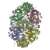
 SCYTALIDIUM THERMOPHILUM (fungus)
 X-RAY DIFFRACTION /
  Authors
 Citation
 Structure visualization
 Downloads & links
 Other downloads


 PDBj
PDBj

 Assembly
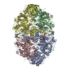


 Mass: 632.487 Da / Num. of mol.: 4 / Source method: obtained synthetically / Formula: C34H32FeN4O5
 Mass: 40.078 Da / Num. of mol.: 9 / Source method: obtained synthetically / Formula: Ca
 Mass: 18.015 Da / Num. of mol.: 2019 / Source method: isolated from a natural source / Formula: H2O
 Sample preparation
 / Beamline: I24 / Wavelength: 0.9686
  Processing