 Movie
Movie Controller


+ Open data

- Basic information
| Entry | Database: PDB / ID: 1sui | ||||||
|---|---|---|---|---|---|---|---|
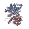
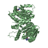
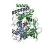
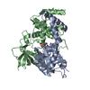
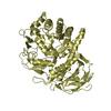
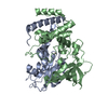
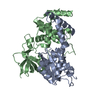
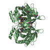
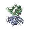
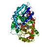
| Title | Alfalfa caffeoyl coenzyme A 3-O-methyltransferase | ||||||
 Components Components | Caffeoyl-CoA O-methyltransferase | ||||||
 Keywords Keywords | TRANSFERASE / ROSSMANN FOLD / PROTEIN-COFACTOR-SUBSTRATE COMPLEX | ||||||
| Function / homology |  Function and homology information Function and homology informationcaffeoyl-CoA O-methyltransferase / caffeoyl-CoA O-methyltransferase activity / lignin biosynthetic process / methylation / metal ion binding Similarity search - Function | ||||||
| Biological species |  | ||||||
| Method |  X-RAY DIFFRACTION / SYNCHROTRON / MAD / Resolution: 2.7 Å X-RAY DIFFRACTION / SYNCHROTRON / MAD / Resolution: 2.7 Å | ||||||
 Authors Authors | Ferrer, J.-L. / Zubieta, C. / Dixon, R.A. / Noel, J.P. | ||||||
 Citation Citation | Journal: Plant Physiol. / Year: 2005 Title: Crystal Structures of Alfalfa Caffeoyl Coenzyme A 3-O-Methyltransferase Authors: Ferrer, J.-L. / Zubieta, C. / Dixon, R.A. / Noel, J.P. | ||||||
| History |
|
- Structure visualization
| Structure viewer | Molecule: MolmilJmol/JSmol |
|---|
- Downloads & links
-Download
| PDBx/mmCIF format | 1sui.cif.gz | 195.5 KB | Display | PDBx/mmCIF format |
|---|---|---|---|---|
| PDB format | pdb1sui.ent.gz | 155.2 KB | Display | PDB format |
| PDBx/mmJSON format | 1sui.json.gz | Tree view | PDBx/mmJSON format | |
| Others |  Other downloads Other downloads |
-Validation report
| Arichive directory | https://data.pdbj.org/pub/pdb/validation_reports/su/1suiftp://data.pdbj.org/pub/pdb/validation_reports/su/1sui | HTTPS FTP |
|---|
-Related structure data

-Links
 PDBj
PDBj- Assembly
| Deposited unit | 
| ||||||||
|---|---|---|---|---|---|---|---|---|---|
| 1 | 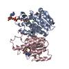
| ||||||||
| 2 | 
| ||||||||
| 3 | 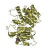
| ||||||||
| Unit cell |
|
-Components
| #1: Protein | Mass: 28036.178 Da / Num. of mol.: 4 Source method: isolated from a genetically manipulated source Source: (gene. exp.)  References: UniProt: Q40313, caffeoyl-CoA O-methyltransferase #2: Chemical | ChemComp-CA /   Mass: 40.078 Da / Num. of mol.: 4 / Source method: obtained synthetically / Formula: Ca Mass: 40.078 Da / Num. of mol.: 4 / Source method: obtained synthetically / Formula: Ca#3: Chemical | ChemComp-SAH /   Type: L-peptide linking / Mass: 384.411 Da / Num. of mol.: 4 / Source method: obtained synthetically / Formula: C14H20N6O5S Type: L-peptide linking / Mass: 384.411 Da / Num. of mol.: 4 / Source method: obtained synthetically / Formula: C14H20N6O5S#4: Chemical | ChemComp-FRE / |   Mass: 959.702 Da / Num. of mol.: 1 / Source method: obtained synthetically / Formula: C31H44N7O20P3S Mass: 959.702 Da / Num. of mol.: 1 / Source method: obtained synthetically / Formula: C31H44N7O20P3S#5: Water | ChemComp-HOH / |  Mass: 18.015 Da / Num. of mol.: 66 / Source method: isolated from a natural source / Formula: H2O Mass: 18.015 Da / Num. of mol.: 66 / Source method: isolated from a natural source / Formula: H2O |
|---|
-Experimental details
-Experiment
| Experiment | Method: X-RAY DIFFRACTION / Number of used crystals: 1 |
|---|
- Sample preparation
| Crystal | Density Matthews: 3.08 Å3/Da / Density % sol: 60.08 % |
|---|---|
| Crystal grow | Temperature: 288 K / pH: 8.5 Details: PEG 8000, TAPS, calcium acetate, pH 8.5, VAPOR DIFFUSION, HANGING DROP, temperature 288K, pH 8.50 |
-Data collection
| Diffraction | Mean temperature: 100 K | ||||||||||||
|---|---|---|---|---|---|---|---|---|---|---|---|---|---|
| Diffraction source | Source: SYNCHROTRON / Site: ESRF  / Beamline: BM30A / Wavelength: 0.97966, 0.97927, 0.97549 / Beamline: BM30A / Wavelength: 0.97966, 0.97927, 0.97549 | ||||||||||||
| Detector | Type: MARRESEARCH / Detector: CCD / Date: Apr 20, 2001 | ||||||||||||
| Radiation | Monochromator: SI (111) / Protocol: MAD / Monochromatic (M) / Laue (L): M / Scattering type: x-ray | ||||||||||||
| Radiation wavelength |
| ||||||||||||
| Reflection | Resolution: 2.7→25 Å / Num. obs: 34905 / % possible obs: 85.7 % / Observed criterion σ(I): -3 / Redundancy: 2.34 % / Biso Wilson estimate: 84.7 Å2 / Rsym value: 0.063 / Net I/σ(I): 22.3 | ||||||||||||
| Reflection shell | Resolution: 2.7→2.78 Å / Redundancy: 1.54 % / Mean I/σ(I) obs: 1 / Rsym value: 0.315 / % possible all: 49.5 |
- Processing
| Software |
| ||||||||||||||||||||||||||||||||||||||||||||||||||||||||||||
|---|---|---|---|---|---|---|---|---|---|---|---|---|---|---|---|---|---|---|---|---|---|---|---|---|---|---|---|---|---|---|---|---|---|---|---|---|---|---|---|---|---|---|---|---|---|---|---|---|---|---|---|---|---|---|---|---|---|---|---|---|---|
| Refinement | Method to determine structure: MAD / Resolution: 2.7→25 Å / Rfactor Rfree error: 0.007 / Isotropic thermal model: ANISOTROPIC / Cross valid method: THROUGHOUT / σ(F): 0 / Stereochemistry target values: MAXIMUM LIKELIHOOD
| ||||||||||||||||||||||||||||||||||||||||||||||||||||||||||||
| Displacement parameters | Biso mean: 84 Å2
| ||||||||||||||||||||||||||||||||||||||||||||||||||||||||||||
| Refine analyze |
| ||||||||||||||||||||||||||||||||||||||||||||||||||||||||||||
| Refinement step | Cycle: LAST / Resolution: 2.7→25 Å
| ||||||||||||||||||||||||||||||||||||||||||||||||||||||||||||
| Refine LS restraints |
| ||||||||||||||||||||||||||||||||||||||||||||||||||||||||||||
| LS refinement shell | Resolution: 2.7→2.73 Å / Total num. of bins used: 6 /
|
