 Movie
Movie Controller

[English] 日本語
 Yorodumi
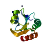
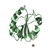
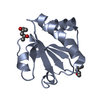
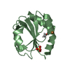
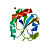
- PDB-1srx: THREE-DIMENSIONAL STRUCTURE OF ESCHERICHIA COLI THIOREDOXIN-S2 TO... -
+ Open data

- Basic information
| Entry | Database: PDB / ID: 1srx | ||||||
|---|---|---|---|---|---|---|---|
| Title | THREE-DIMENSIONAL STRUCTURE OF ESCHERICHIA COLI THIOREDOXIN-S2 TO 2.8 ANGSTROMS RESOLUTION | ||||||
 Components Components | THIOREDOXIN | ||||||
 Keywords Keywords | ELECTRON TRANSPORT | ||||||
| Function / homology |  Function and homology information Function and homology informationDNA polymerase processivity factor activity / protein-disulfide reductase activity / cell redox homeostasis / cytoplasm / cytosol Similarity search - Function | ||||||
| Biological species |  | ||||||
| Method |  X-RAY DIFFRACTION / Resolution: 2.8 Å X-RAY DIFFRACTION / Resolution: 2.8 Å | ||||||
 Authors Authors | Soderberg, B.-O. | ||||||
 Citation Citation | Journal: Proc.Natl.Acad.Sci.USA / Year: 1975 Title: Three-dimensional structure of Escherichia coli thioredoxin-S2 to 2.8 A resolution. Authors: Holmgren, A. / Soderberg, B.O. / Eklund, H. / Branden, C.I. #1: Journal: J.Mol.Biol. / Year: 1974Title: Structure of Oxidized Thioredoxin to 4.5 Angstroms Resolution Authors: Soderberg, B.-O. / Holmgren, A. / Branden, C.-I. | ||||||
| History |
|
- Structure visualization
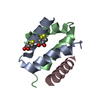
| Structure viewer | Molecule: MolmilJmol/JSmol |
|---|
- Downloads & links
-Download
| PDBx/mmCIF format | 1srx.cif.gz | 13.5 KB | Display | PDBx/mmCIF format |
|---|---|---|---|---|
| PDB format | pdb1srx.ent.gz | 5.7 KB | Display | PDB format |
| PDBx/mmJSON format | 1srx.json.gz | Tree view | PDBx/mmJSON format | |
| Others |  Other downloads Other downloads |
-Validation report
| Arichive directory | https://data.pdbj.org/pub/pdb/validation_reports/sr/1srxftp://data.pdbj.org/pub/pdb/validation_reports/sr/1srx | HTTPS FTP |
|---|
-Related structure data
| Similar structure data |
|---|


-Links
 PDBj
PDBj

- Assembly
| Deposited unit | 
| ||||||||
|---|---|---|---|---|---|---|---|---|---|
| 1 |
| ||||||||
| Unit cell |
|
-Components
| #1: Protein | Mass: 11759.448 Da / Num. of mol.: 1 Source method: isolated from a genetically manipulated source Source: (gene. exp.) |
|---|---|
| Compound details | THE ACTIVE SITE DISULFIDE BOND IS BETWEEN CYS-32 AND CYS-35 SITE S-S IS THE ACTIVE CENTER DISULFIDE ...THE ACTIVE SITE DISULFIDE BOND IS BETWEEN CYS-32 AND CYS-35 SITE S-S IS THE ACTIVE CENTER DISULFIDE BRIDGED REVERSE TURN. RESIDUES OTHER THAN THOSE GIVEN UNDER SITE BELOW MAY BE INVOLVED IN THE ACTIVITY. |
-Experimental details
-Experiment
| Experiment | Method: X-RAY DIFFRACTION |
|---|
- Sample preparation
| Crystal | Density Matthews: 5.39 Å3/Da / Density % sol: 77.17 % |
|---|---|
| Crystal grow | *PLUS Method: other / Details: Soderberg, B.O., (1974) J. Mol. Biol., 90, 143. |
- Processing
| Refinement | Highest resolution: 2.8 Å | ||||||||||||
|---|---|---|---|---|---|---|---|---|---|---|---|---|---|
| Refinement step | Cycle: LAST / Highest resolution: 2.8 Å
|
