 Movie
Movie Controller

[English] 日本語
 Yorodumi
- PDB-1sq7: Understanding protein lids: Structural analysis of active hinge m... -
+ Open data

- Basic information
| Entry | Database: PDB / ID: 1sq7 | ||||||
|---|---|---|---|---|---|---|---|
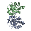
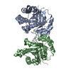
| Title | Understanding protein lids: Structural analysis of active hinge mutants in triosephosphate isomerase | ||||||
 Components Components | Triosephosphate isomerase | ||||||
 Keywords Keywords | ISOMERASE / archae / evolution / flexible loop-6 / TIM / N-hinge | ||||||
| Function / homology |  Function and homology information Function and homology informationGlycolysis / Glycolysis / Gluconeogenesis / Gluconeogenesis / methylglyoxal biosynthetic process / methylglyoxal synthase / methylglyoxal synthase activity / triose-phosphate isomerase / triose-phosphate isomerase activity / glyceraldehyde-3-phosphate biosynthetic process ...Glycolysis / Glycolysis / Gluconeogenesis / Gluconeogenesis / methylglyoxal biosynthetic process / methylglyoxal synthase / methylglyoxal synthase activity / triose-phosphate isomerase / triose-phosphate isomerase activity / glyceraldehyde-3-phosphate biosynthetic process / glycerol catabolic process / canonical glycolysis / gluconeogenesis / glycolytic process / ubiquitin protein ligase binding / protein homodimerization activity / cytosol Similarity search - Function | ||||||
| Biological species |  | ||||||
| Method |  X-RAY DIFFRACTION / SYNCHROTRON / MOLECULAR REPLACEMENT / Resolution: 2.85 Å X-RAY DIFFRACTION / SYNCHROTRON / MOLECULAR REPLACEMENT / Resolution: 2.85 Å | ||||||
 Authors Authors | Kursula, I. / Salin, M. / Sun, J. / Norledge, B.V. / Haapalainen, A.M. / Sampson, N.S. / Wierenga, R.K. | ||||||
 Citation Citation | Journal: Protein Eng.Des.Sel. / Year: 2004 Title: Understanding protein lids: structural analysis of active hinge mutants in triosephosphate isomerase Authors: Kursula, I. / Salin, M. / Sun, J. / Norledge, B.V. / Haapalainen, A.M. / Sampson, N.S. / Wierenga, R.K. | ||||||
| History |
|
- Structure visualization
| Structure viewer | Molecule: MolmilJmol/JSmol |
|---|
- Downloads & links
-Download
| PDBx/mmCIF format | 1sq7.cif.gz | 104.3 KB | Display | PDBx/mmCIF format |
|---|---|---|---|---|
| PDB format | pdb1sq7.ent.gz | 80.4 KB | Display | PDB format |
| PDBx/mmJSON format | 1sq7.json.gz | Tree view | PDBx/mmJSON format | |
| Others |  Other downloads Other downloads |
-Validation report
| Arichive directory | https://data.pdbj.org/pub/pdb/validation_reports/sq/1sq7ftp://data.pdbj.org/pub/pdb/validation_reports/sq/1sq7 | HTTPS FTP |
|---|
-Related structure data
| Related structure data |  1spqC  1ssdC  1ssgC  1su5C  1sw0C  1sw3C  1sw7C  8timS C: citing same article ( S: Starting model for refinement |
|---|---|
| Similar structure data |


-Links
 PDBj
PDBj
- Assembly
| Deposited unit | 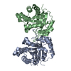
| ||||||||
|---|---|---|---|---|---|---|---|---|---|
| 1 |
| ||||||||
| Unit cell |
| ||||||||
| Components on special symmetry positions |
| ||||||||
| Details | asymmetric unit contains one biological unit, dimer (chains A and B) |
-Components
| #1: Protein | Mass: 26595.348 Da / Num. of mol.: 2 / Mutation: K174L, T175W Source method: isolated from a genetically manipulated source Source: (gene. exp.)  #2: Water | ChemComp-HOH / |  Mass: 18.015 Da / Num. of mol.: 211 / Source method: isolated from a natural source / Formula: H2O Mass: 18.015 Da / Num. of mol.: 211 / Source method: isolated from a natural source / Formula: H2O |
|---|
-Experimental details
-Experiment
| Experiment | Method: X-RAY DIFFRACTION / Number of used crystals: 1 |
|---|
- Sample preparation
| Crystal | Density Matthews: 2.58 Å3/Da / Density % sol: 52.4 % |
|---|---|
| Crystal grow | Temperature: 295 K / Method: vapor diffusion, hanging drop / pH: 5.2 Details: PEG 6000, citrate, t-butanol, pH 5.2, VAPOR DIFFUSION, HANGING DROP, temperature 295K |
-Data collection
| Diffraction | Mean temperature: 100 K |
|---|---|
| Diffraction source | Source: SYNCHROTRON / Site: EMBL/DESY, HAMBURG  / Beamline: X11 / Wavelength: 0.8 Å / Beamline: X11 / Wavelength: 0.8 Å |
| Detector | Type: MARRESEARCH / Detector: CCD / Date: Jun 15, 2002 / Details: Bent mirror |
| Radiation | Monochromator: Triangular / Protocol: SINGLE WAVELENGTH / Monochromatic (M) / Laue (L): M / Scattering type: x-ray |
| Radiation wavelength | Wavelength: 0.8 Å / Relative weight: 1 |
| Reflection | Resolution: 2.8→30 Å / Num. all: 16236 / Num. obs: 15311 / % possible obs: 94.3 % / Observed criterion σ(F): 0 / Observed criterion σ(I): 0 / Redundancy: 6.62 % / Biso Wilson estimate: 20.4 Å2 / Rmerge(I) obs: 0.083 / Net I/σ(I): 10.42 |
| Reflection shell | Resolution: 2.76→2.93 Å / Redundancy: 5.29 % / Rmerge(I) obs: 0.117 / Mean I/σ(I) obs: 7 / Num. unique all: 2572 / % possible all: 82.4 |
- Processing
| Software |
| ||||||||||||||||||||||||||||||||||||||||||||||||||||||||||||||||||||||||||||||||||||||||||||||||||||
|---|---|---|---|---|---|---|---|---|---|---|---|---|---|---|---|---|---|---|---|---|---|---|---|---|---|---|---|---|---|---|---|---|---|---|---|---|---|---|---|---|---|---|---|---|---|---|---|---|---|---|---|---|---|---|---|---|---|---|---|---|---|---|---|---|---|---|---|---|---|---|---|---|---|---|---|---|---|---|---|---|---|---|---|---|---|---|---|---|---|---|---|---|---|---|---|---|---|---|---|---|---|
| Refinement | Method to determine structure: MOLECULAR REPLACEMENT Starting model: PDB entry 8TIM Resolution: 2.85→28.28 Å / Cor.coef. Fo:Fc: 0.911 / Cor.coef. Fo:Fc free: 0.875 / SU B: 14.334 / SU ML: 0.274 / TLS residual ADP flag: LIKELY RESIDUAL / Cross valid method: THROUGHOUT / σ(F): 0 / σ(I): 0 / ESU R Free: 0.399 / Stereochemistry target values: MAXIMUM LIKELIHOOD / Details: HYDROGENS HAVE BEEN ADDED IN THE RIDING POSITIONS
| ||||||||||||||||||||||||||||||||||||||||||||||||||||||||||||||||||||||||||||||||||||||||||||||||||||
| Solvent computation | Ion probe radii: 0.8 Å / Shrinkage radii: 0.8 Å / VDW probe radii: 1.4 Å / Solvent model: BABINET MODEL WITH MASK | ||||||||||||||||||||||||||||||||||||||||||||||||||||||||||||||||||||||||||||||||||||||||||||||||||||
| Displacement parameters | Biso mean: 3.209 Å2
| ||||||||||||||||||||||||||||||||||||||||||||||||||||||||||||||||||||||||||||||||||||||||||||||||||||
| Refinement step | Cycle: LAST / Resolution: 2.85→28.28 Å
| ||||||||||||||||||||||||||||||||||||||||||||||||||||||||||||||||||||||||||||||||||||||||||||||||||||
| Refine LS restraints |
| ||||||||||||||||||||||||||||||||||||||||||||||||||||||||||||||||||||||||||||||||||||||||||||||||||||
| LS refinement shell | Resolution: 2.85→2.923 Å / Total num. of bins used: 20
| ||||||||||||||||||||||||||||||||||||||||||||||||||||||||||||||||||||||||||||||||||||||||||||||||||||
| Refinement TLS params. | Method: refined / Refine-ID: X-RAY DIFFRACTION
| ||||||||||||||||||||||||||||||||||||||||||||||||||||||||||||||||||||||||||||||||||||||||||||||||||||
| Refinement TLS group |
|
