 Movie
Movie Controller

[English] 日本語
 Yorodumi
- PDB-1son: ADENYLOSUCCINATE SYNTHETASE IN COMPLEX WITH THE NATURAL FEEDBACK ... -
+ Open data

- Basic information
| Entry | Database: PDB / ID: 1son | ||||||
|---|---|---|---|---|---|---|---|
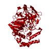
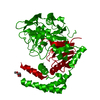
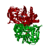
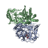
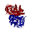
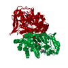
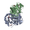
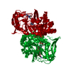
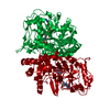
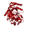
| Title | ADENYLOSUCCINATE SYNTHETASE IN COMPLEX WITH THE NATURAL FEEDBACK INHIBITOR AMP | ||||||
 Components Components | ADENYLOSUCCINATE SYNTHETASE | ||||||
 Keywords Keywords | LIGASE / PURINE NUCLEOTIDE BIOSYNTHESIS / GTP-HYDROLYZING ENZYME / HERBICIDE / SYNTHETASE | ||||||
| Function / homology |  Function and homology information Function and homology informationadenylosuccinate synthase / adenylosuccinate synthase activity / adenosine biosynthetic process / IMP metabolic process / 'de novo' AMP biosynthetic process / nucleobase-containing small molecule interconversion / purine nucleotide biosynthetic process / guanosine tetraphosphate binding / DNA damage response / GTP binding ...adenylosuccinate synthase / adenylosuccinate synthase activity / adenosine biosynthetic process / IMP metabolic process / 'de novo' AMP biosynthetic process / nucleobase-containing small molecule interconversion / purine nucleotide biosynthetic process / guanosine tetraphosphate binding / DNA damage response / GTP binding / magnesium ion binding / membrane / cytoplasm / cytosol Similarity search - Function | ||||||
| Biological species |  | ||||||
| Method |  X-RAY DIFFRACTION / RIGID BODY REFINEMENT / Resolution: 2.55 Å X-RAY DIFFRACTION / RIGID BODY REFINEMENT / Resolution: 2.55 Å | ||||||
 Authors Authors | Cowan-Jacob, S.W. | ||||||
 Citation Citation | Journal: Proc.Natl.Acad.Sci.USA / Year: 1996 Title: The mode of action and the structure of a herbicide in complex with its target: binding of activated hydantocidin to the feedback regulation site of adenylosuccinate synthetase. Authors: Fonne-Pfister, R. / Chemla, P. / Ward, E. / Girardet, M. / Kreuz, K.E. / Honzatko, R.B. / Fromm, H.J. / Schar, H.P. / Grutter, M.G. / Cowan-Jacob, S.W. #1: Journal: J.Mol.Biol. / Year: 1995Title: Refined Crystal Structures of Unligated Adenylosuccinate Synthetase from Escherichia Coli Authors: Silva, M.M. / Poland, B.W. / Hoffman, C.R. / Fromm, H.J. / Honzatko, R.B. #2: Journal: J.Biol.Chem. / Year: 1993Title: Crystal Structure of Adenylosuccinate Synthetase from Escherichia Coli. Evidence for Convergent Evolution of GTP-Binding Domains Authors: Poland, B.W. / Silva, M.M. / Serra, M.A. / Cho, Y. / Kim, K.H. / Harris, E.M. / Honzatko, R.B. #3: Journal: J.Mol.Biol. / Year: 1988Title: Preliminary X-Ray Crystallographic Study of Adenylosuccinate Synthetase from Escherichia Coli Authors: Serra, M.A. / Bass, M.B. / Fromm, H.J. / Honzatko, R.B. | ||||||
| History |
|
- Structure visualization
| Structure viewer | Molecule: MolmilJmol/JSmol |
|---|
- Downloads & links
-Download
| PDBx/mmCIF format | 1son.cif.gz | 85.9 KB | Display | PDBx/mmCIF format |
|---|---|---|---|---|
| PDB format | pdb1son.ent.gz | 68.9 KB | Display | PDB format |
| PDBx/mmJSON format | 1son.json.gz | Tree view | PDBx/mmJSON format | |
| Others |  Other downloads Other downloads |
-Validation report
| Arichive directory | https://data.pdbj.org/pub/pdb/validation_reports/so/1sonftp://data.pdbj.org/pub/pdb/validation_reports/so/1son | HTTPS FTP |
|---|
-Related structure data
| Related structure data |  1sooSC S: Starting model for refinement C: citing same article ( |
|---|---|
| Similar structure data |
-Links
 PDBj
PDBj


- Assembly
| Deposited unit | 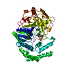
| ||||||||
|---|---|---|---|---|---|---|---|---|---|
| 1 | 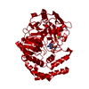
| ||||||||
| Unit cell |
| ||||||||
| Components on special symmetry positions |
|
-Components
| #1: Protein | Mass: 47269.598 Da / Num. of mol.: 1 / Source method: isolated from a natural source / Source: (natural) |
|---|---|
| #2: Chemical | ChemComp-AMP /   Mass: 347.221 Da / Num. of mol.: 1 / Source method: obtained synthetically / Formula: C10H14N5O7P / Comment: AMP*YM Mass: 347.221 Da / Num. of mol.: 1 / Source method: obtained synthetically / Formula: C10H14N5O7P / Comment: AMP*YM |
| #3: Chemical | ChemComp-BME /   Mass: 78.133 Da / Num. of mol.: 1 / Source method: obtained synthetically / Formula: C2H6OS Mass: 78.133 Da / Num. of mol.: 1 / Source method: obtained synthetically / Formula: C2H6OS |
| #4: Water | ChemComp-HOH /  Mass: 18.015 Da / Num. of mol.: 93 / Source method: isolated from a natural source / Formula: H2O Mass: 18.015 Da / Num. of mol.: 93 / Source method: isolated from a natural source / Formula: H2O |
-Experimental details
-Experiment
| Experiment | Method: X-RAY DIFFRACTION / Number of used crystals: 1 |
|---|
- Sample preparation
| Crystal | Density Matthews: 2.65 Å3/Da / Density % sol: 54 % | ||||||||||||||||||||||||||||||||||||||||||
|---|---|---|---|---|---|---|---|---|---|---|---|---|---|---|---|---|---|---|---|---|---|---|---|---|---|---|---|---|---|---|---|---|---|---|---|---|---|---|---|---|---|---|---|
| Crystal grow | pH: 8.6 / Details: pH 8.6 | ||||||||||||||||||||||||||||||||||||||||||
| Crystal grow | *PLUS Temperature: 20-25 ℃ / pH: 7.7 / Method: vapor diffusion | ||||||||||||||||||||||||||||||||||||||||||
| Components of the solutions | *PLUS
|
-Data collection
| Diffraction | Mean temperature: 298 K |
|---|---|
| Diffraction source | Wavelength: 1.5418 |
| Detector | Type: MARRESEARCH / Detector: IMAGE PLATE / Date: Oct 13, 1994 |
| Radiation | Monochromatic (M) / Laue (L): M / Scattering type: x-ray |
| Radiation wavelength | Wavelength: 1.5418 Å / Relative weight: 1 |
| Reflection | Resolution: 2.55→28 Å / Num. obs: 17293 / % possible obs: 99.7 % / Observed criterion σ(I): 0 / Redundancy: 6.7 % / Rmerge(I) obs: 0.086 |
| Reflection shell | Resolution: 2.55→2.62 Å / Redundancy: 6.3 % / Rmerge(I) obs: 0.4 / % possible all: 99.2 |
| Reflection shell | *PLUS % possible obs: 99.2 % |
- Processing
| Software |
| ||||||||||||||||||||||||||||||||||||||||||||||||||||||||||||
|---|---|---|---|---|---|---|---|---|---|---|---|---|---|---|---|---|---|---|---|---|---|---|---|---|---|---|---|---|---|---|---|---|---|---|---|---|---|---|---|---|---|---|---|---|---|---|---|---|---|---|---|---|---|---|---|---|---|---|---|---|---|
| Refinement | Method to determine structure: RIGID BODY REFINEMENT Starting model: 1SOO Resolution: 2.55→8 Å / σ(F): 0
| ||||||||||||||||||||||||||||||||||||||||||||||||||||||||||||
| Displacement parameters | Biso mean: 34.2 Å2 | ||||||||||||||||||||||||||||||||||||||||||||||||||||||||||||
| Refine analyze | Luzzati coordinate error obs: 0.4 Å | ||||||||||||||||||||||||||||||||||||||||||||||||||||||||||||
| Refinement step | Cycle: LAST / Resolution: 2.55→8 Å
| ||||||||||||||||||||||||||||||||||||||||||||||||||||||||||||
| Refine LS restraints |
| ||||||||||||||||||||||||||||||||||||||||||||||||||||||||||||
| Software | *PLUS Name: X-PLOR / Version: 3.1 / Classification: refinement | ||||||||||||||||||||||||||||||||||||||||||||||||||||||||||||
| Refinement | *PLUS | ||||||||||||||||||||||||||||||||||||||||||||||||||||||||||||
| Solvent computation | *PLUS | ||||||||||||||||||||||||||||||||||||||||||||||||||||||||||||
| Displacement parameters | *PLUS | ||||||||||||||||||||||||||||||||||||||||||||||||||||||||||||
| Refine LS restraints | *PLUS
|
