 Movie
Movie Controller

[English] 日本語
 Yorodumi
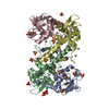
- PDB-1sms: Structure of the Ribonucleotide Reductase Rnr4 Homodimer from Sac... -
+ Open data

- Basic information
| Entry | Database: PDB / ID: 1sms | ||||||
|---|---|---|---|---|---|---|---|
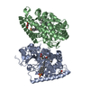
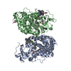
| Title | Structure of the Ribonucleotide Reductase Rnr4 Homodimer from Saccharomyces cerevisiae | ||||||
 Components Components | Ribonucleoside-diphosphate reductase small chain 2 | ||||||
 Keywords Keywords | OXIDOREDUCTASE | ||||||
| Function / homology |  Function and homology information Function and homology informationInterconversion of nucleotide di- and triphosphates / ribonucleoside-diphosphate reductase complex / ribonucleoside-diphosphate reductase / ribonucleoside-diphosphate reductase activity, thioredoxin disulfide as acceptor / deoxyribonucleotide biosynthetic process / protein heterodimerization activity / nucleus / cytoplasm Similarity search - Function | ||||||
| Biological species |  | ||||||
| Method |  X-RAY DIFFRACTION / SYNCHROTRON / MOLECULAR REPLACEMENT / Resolution: 3.1 Å X-RAY DIFFRACTION / SYNCHROTRON / MOLECULAR REPLACEMENT / Resolution: 3.1 Å | ||||||
 Authors Authors | Sommerhalter, M. / Voegtli, W.C. / Perlstein, D.L. / Ge, J. / Stubbe, J. / Rosenzweig, A.C. | ||||||
 Citation Citation | Journal: Biochemistry / Year: 2004 Title: Structures of the yeast ribonucleotide reductase Rnr2 and Rnr4 homodimers. Authors: Sommerhalter, M. / Voegtli, W.C. / Perlstein, D.L. / Ge, J. / Stubbe, J. / Rosenzweig, A.C. | ||||||
| History |
|
- Structure visualization
| Structure viewer | Molecule: MolmilJmol/JSmol |
|---|
- Downloads & links
-Download
| PDBx/mmCIF format | 1sms.cif.gz | 138.3 KB | Display | PDBx/mmCIF format |
|---|---|---|---|---|
| PDB format | pdb1sms.ent.gz | 108.6 KB | Display | PDB format |
| PDBx/mmJSON format | 1sms.json.gz | Tree view | PDBx/mmJSON format | |
| Others |  Other downloads Other downloads |
-Validation report
| Arichive directory | https://data.pdbj.org/pub/pdb/validation_reports/sm/1smsftp://data.pdbj.org/pub/pdb/validation_reports/sm/1sms | HTTPS FTP |
|---|
-Related structure data
| Related structure data |  1smqC 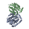 1jk0S S: Starting model for refinement C: citing same article ( |
|---|---|
| Similar structure data |


-Links
 PDBj
PDBj


- Assembly
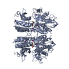
| Deposited unit | 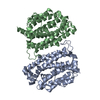
| ||||||||
|---|---|---|---|---|---|---|---|---|---|
| 1 |
| ||||||||
| Unit cell |
|
-Components
| #1: Protein | Mass: 40102.500 Da / Num. of mol.: 2 Source method: isolated from a genetically manipulated source Source: (gene. exp.) Gene: RNR4, YGR180C / Production host:  References: UniProt: P49723, ribonucleoside-diphosphate reductase #2: Chemical | ChemComp-HG /   Mass: 200.590 Da / Num. of mol.: 4 / Source method: obtained synthetically / Formula: Hg Mass: 200.590 Da / Num. of mol.: 4 / Source method: obtained synthetically / Formula: Hg |
|---|
-Experimental details
-Experiment
| Experiment | Method: X-RAY DIFFRACTION / Number of used crystals: 1 |
|---|
- Sample preparation
| Crystal | Density Matthews: 2.49 Å3/Da / Density % sol: 50.53 % |
|---|---|
| Crystal grow | Temperature: 298 K / Method: vapor diffusion, hanging drop / pH: 7.5 Details: HEPES, PEG 1000, Ethanol, sodium chloride, EMTS, pH 7.5, VAPOR DIFFUSION, HANGING DROP, temperature 298K |
-Data collection
| Diffraction | Mean temperature: 113 K |
|---|---|
| Diffraction source | Source: SYNCHROTRON / Site: APS  / Beamline: 5ID-B / Wavelength: 1 Å / Beamline: 5ID-B / Wavelength: 1 Å |
| Detector | Type: MARRESEARCH / Detector: CCD |
| Radiation | Protocol: SINGLE WAVELENGTH / Monochromatic (M) / Laue (L): M / Scattering type: x-ray |
| Radiation wavelength | Wavelength: 1 Å / Relative weight: 1 |
| Reflection | Resolution: 3.1→12 Å / Num. obs: 239131 / % possible obs: 93.6 % / Rsym value: 0.088 / Net I/σ(I): 13.4 |
| Reflection shell | Resolution: 3.1→3.15 Å / Mean I/σ(I) obs: 2.4 / Rsym value: 0.424 / % possible all: 94.1 |
- Processing
| Software |
| ||||||||||||||||
|---|---|---|---|---|---|---|---|---|---|---|---|---|---|---|---|---|---|
| Refinement | Method to determine structure: MOLECULAR REPLACEMENT Starting model: PDB entry 1JK0 subunit Y4 Resolution: 3.1→12 Å / Cross valid method: THROUGHOUT
| ||||||||||||||||
| Refinement step | Cycle: LAST / Resolution: 3.1→12 Å
| ||||||||||||||||
| Refine LS restraints |
|
