 ムービー
ムービー コントローラー


+ データを開く

- 基本情報
基本情報
| 登録情報 | データベース: PDB / ID: 1sh5 | ||||||
|---|---|---|---|---|---|---|---|
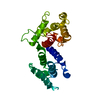
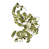
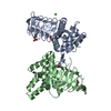
| タイトル | Crystal structure of actin-binding domain of mouse plectin | ||||||
 要素 要素 | Plectin 1 | ||||||
 キーワード キーワード | STRUCTURAL PROTEIN / plectin / actin-binding domain / calponin-homology domain | ||||||
| 機能・相同性 |  機能・相同性情報 機能・相同性情報Type I hemidesmosome assembly / protein-containing complex organization / actomyosin contractile ring assembly actin filament organization / Caspase-mediated cleavage of cytoskeletal proteins / tight junction organization / skeletal myofibril assembly / hemidesmosome assembly / hemidesmosome / contractile muscle fiber / leukocyte migration involved in immune response ...Type I hemidesmosome assembly / protein-containing complex organization / actomyosin contractile ring assembly actin filament organization / Caspase-mediated cleavage of cytoskeletal proteins / tight junction organization / skeletal myofibril assembly / hemidesmosome assembly / hemidesmosome / contractile muscle fiber / leukocyte migration involved in immune response / intermediate filament organization / intermediate filament cytoskeleton organization / dystroglycan binding / fibroblast migration / cellular response to hydrostatic pressure / adherens junction organization / regulation of vascular permeability / myelination in peripheral nervous system / T cell chemotaxis / cellular response to fluid shear stress / peripheral nervous system myelin maintenance / intermediate filament cytoskeleton / myoblast differentiation / cardiac muscle cell development / podosome / ankyrin binding / sarcomere organization / response to food / skin development / nucleus organization / keratinocyte development / myofibril / transmission of nerve impulse / brush border / establishment of skin barrier / skeletal muscle tissue development / keratinocyte differentiation / skeletal muscle fiber development / actin filament organization / cell periphery / respiratory electron transport chain / mitochondrion organization / cell motility / cellular response to mechanical stimulus / sarcolemma / multicellular organism growth / Z disc / cell morphogenesis / actin filament binding / intracellular protein localization / myelin sheath / actin binding / actin cytoskeleton organization / gene expression / mitochondrial outer membrane / axon / focal adhesion / dendrite / identical protein binding / cytoplasm / cytosol 類似検索 - 分子機能 | ||||||
| 生物種 |  | ||||||
| 手法 |  X線回折 / シンクロトロン / 分子置換 / 解像度: 2 Å X線回折 / シンクロトロン / 分子置換 / 解像度: 2 Å | ||||||
 データ登録者 データ登録者 | Sevcik, J. / Urbanikova, L. | ||||||
 引用 引用 | ジャーナル: Eur.J.Biochem. / 年: 2004 タイトル: Actin-binding domain of mouse plectin: crystal structure and binding to vimentin 著者: Sevcik, J. / Urbanikova, L. / Kostan, J. / Janda, L. / Wiche, G. #1: ジャーナル: Acta Crystallogr.,Sect.D / 年: 2002タイトル: Purification, crystallization and preliminary X-ray analysis of the plectin actin-binding domain 著者: Urbanikova, L. / Janda, L. / Popov, A. / Wiche, G. / Sevcik, J. | ||||||
| 履歴 |
|
- 構造の表示
構造の表示
| 構造ビューア | 分子: MolmilJmol/JSmol |
|---|
- ダウンロードとリンク
-ダウンロード
| PDBx/mmCIF形式 | 1sh5.cif.gz | 292.4 KB | 表示 | PDBx/mmCIF形式 |
|---|---|---|---|---|
| PDB形式 | pdb1sh5.ent.gz | 244.9 KB | 表示 | PDB形式 |
| PDBx/mmJSON形式 | 1sh5.json.gz | ツリー表示 | PDBx/mmJSON形式 | |
| その他 |  その他のダウンロード その他のダウンロード |
-検証レポート
| 文書・要旨 | 1sh5_validation.pdf.gz | 428.1 KB | 表示 | wwPDB検証レポート |
|---|---|---|---|---|
| 文書・詳細版 | 1sh5_full_validation.pdf.gz | 444.6 KB | 表示 | |
| XML形式データ | 1sh5_validation.xml.gz | 21.8 KB | 表示 | |
| CIF形式データ | 1sh5_validation.cif.gz | 30.5 KB | 表示 | |
| アーカイブディレクトリ | https://data.pdbj.org/pub/pdb/validation_reports/sh/1sh5ftp://data.pdbj.org/pub/pdb/validation_reports/sh/1sh5 | HTTPS FTP |
-関連構造データ


-リンク
 PDBj
PDBj
- 集合体
集合体
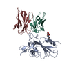
| 登録構造単位 | 
| ||||||||||
|---|---|---|---|---|---|---|---|---|---|---|---|
| 1 |
| ||||||||||
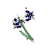
| 単位格子 |
|
-要素
| #1: タンパク質 | 分子量: 28717.717 Da / 分子数: 2 / 断片: actin-binding domain / 由来タイプ: 組換発現 / 由来: (組換発現)  #2: 水 | ChemComp-HOH / |  分子量: 18.015 Da / 分子数: 188 / 由来タイプ: 天然 / 式: H2O 分子量: 18.015 Da / 分子数: 188 / 由来タイプ: 天然 / 式: H2O |
|---|
-実験情報
-実験
| 実験 | 手法: X線回折 / 使用した結晶の数: 1 |
|---|
- 試料調製
試料調製
| 結晶 | マシュー密度: 3.08 Å3/Da / 溶媒含有率: 59.8 % |
|---|---|
| 結晶化 | 温度: 298 K / 手法: 蒸気拡散法, ハンギングドロップ法 / pH: 8.5 詳細: PEG 8000, Tris-HCl, pH 8.5, VAPOR DIFFUSION, HANGING DROP, temperature 298K |
-データ収集
| 回折 | 平均測定温度: 293 K |
|---|---|
| 放射光源 | 由来: シンクロトロン / サイト: EMBL/DESY, HAMBURG  / ビームライン: X31 / 波長: 1.1 Å / ビームライン: X31 / 波長: 1.1 Å |
| 検出器 | タイプ: MARRESEARCH / 検出器: IMAGE PLATE / 日付: 2000年9月16日 / 詳細: mirrors |
| 放射 | モノクロメーター: Si111 / プロトコル: SINGLE WAVELENGTH / 単色(M)・ラウエ(L): M / 散乱光タイプ: x-ray |
| 放射波長 | 波長: 1.1 Å / 相対比: 1 |
| 反射 | 解像度: 1.95→30 Å / Num. all: 49742 / Num. obs: 47802 / % possible obs: 96.2 % / 冗長度: 3.5 % / Biso Wilson estimate: 32.6 Å2 / Rmerge(I) obs: 0.06 / Net I/σ(I): 22.2 |
| 反射 シェル | 解像度: 1.95→1.97 Å / 冗長度: 2.1 % / Rmerge(I) obs: 0.4 / Mean I/σ(I) obs: 2.1 / Num. unique all: 1187 / % possible all: 71.7 |
- 解析
解析
| ソフトウェア |
| |||||||||||||||||||||||||||||||||||||||||||||||||||||||||||||||||||||||||||||||||||||||||||||||||||||||||||||||||||
|---|---|---|---|---|---|---|---|---|---|---|---|---|---|---|---|---|---|---|---|---|---|---|---|---|---|---|---|---|---|---|---|---|---|---|---|---|---|---|---|---|---|---|---|---|---|---|---|---|---|---|---|---|---|---|---|---|---|---|---|---|---|---|---|---|---|---|---|---|---|---|---|---|---|---|---|---|---|---|---|---|---|---|---|---|---|---|---|---|---|---|---|---|---|---|---|---|---|---|---|---|---|---|---|---|---|---|---|---|---|---|---|---|---|---|---|---|
| 精密化 | 構造決定の手法: 分子置換 開始モデル: PDB ENTRY 1QAG 解像度: 2→30 Å / Cor.coef. Fo:Fc: 0.974 / Cor.coef. Fo:Fc free: 0.957 / SU B: 2.892 / SU ML: 0.081 / 交差検証法: THROUGHOUT / ESU R: 0.242 / ESU R Free: 0.122 / 立体化学のターゲット値: MAXIMUM LIKELIHOOD / 詳細: HYDROGENS HAVE BEEN ADDED IN THE RIDING POSITIONS
| |||||||||||||||||||||||||||||||||||||||||||||||||||||||||||||||||||||||||||||||||||||||||||||||||||||||||||||||||||
| 溶媒の処理 | イオンプローブ半径: 0.8 Å / 減衰半径: 0.8 Å / VDWプローブ半径: 1.4 Å / 溶媒モデル: BABINET MODEL WITH MASK | |||||||||||||||||||||||||||||||||||||||||||||||||||||||||||||||||||||||||||||||||||||||||||||||||||||||||||||||||||
| 原子変位パラメータ | Biso mean: 39.942 Å2
| |||||||||||||||||||||||||||||||||||||||||||||||||||||||||||||||||||||||||||||||||||||||||||||||||||||||||||||||||||
| 精密化ステップ | サイクル: LAST / 解像度: 2→30 Å
| |||||||||||||||||||||||||||||||||||||||||||||||||||||||||||||||||||||||||||||||||||||||||||||||||||||||||||||||||||
| 拘束条件 |
| |||||||||||||||||||||||||||||||||||||||||||||||||||||||||||||||||||||||||||||||||||||||||||||||||||||||||||||||||||
| LS精密化 シェル | 解像度: 2→2.052 Å / Total num. of bins used: 20
|
