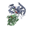
- PDB-1sg9: Crystal structure of Thermotoga maritima protein HEMK, an N5-glut... -
+
Open data
ID or keywords:
Loading...
-
Basic information
Entry
Database: PDB / ID: 1sg9
Title
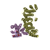
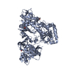
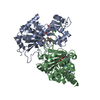
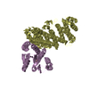
Crystal structure of Thermotoga maritima protein HEMK, an N5-glutamine methyltransferase
Components
hemK protein
Keywords
UNKNOWN FUNCTION / structural genomics / protein structure initiative / HEMK protein / hypothetical protein / PSI / New York SGX Research Center for Structural Genomics / NYSGXRC
Monochromator: Graphite / Protocol: SINGLE WAVELENGTH / Monochromatic (M) / Laue (L): M / Scattering type: x-ray
Radiation wavelength
Wavelength: 0.95 Å / Relative weight: 1
Reflection
Resolution: 2.3→33.09 Å / Num. all: 68970 / Num. obs: 63152 / % possible obs: 89.1 % / Observed criterion σ(F): 0 / Redundancy: 8 % / Biso Wilson estimate: 21.7 Å2 / Rmerge(I) obs: 0.071 / Net I/σ(I): 14
Reflection shell
Resolution: 2.3→2.38 Å / Rmerge(I) obs: 0.622 / % possible all: 90.7
-
Processing
Software
Name
Version
Classification
CNS
1.1
refinement
MARMAD
datareduction
ADSC
datacollection
SCALEPACK
datascaling
AMoRE
phasing
Refinement
Method to determine structure: MOLECULAR REPLACEMENT / Resolution: 2.3→33.09 Å / Rfactor Rfree error: 0.006 / Data cutoff high absF: 192461.58 / Data cutoff low absF: 0 / Isotropic thermal model: RESTRAINED / Cross valid method: THROUGHOUT / σ(F): 0 Details: The electron density suggests that monomers A and C form a dimer via glutamate anhydride, while Monomer B forms a similar dimer with its symmetry related molecule (see remark 350). The ...Details: The electron density suggests that monomers A and C form a dimer via glutamate anhydride, while Monomer B forms a similar dimer with its symmetry related molecule (see remark 350). The glutamate residues (Glu 97 in all chains) involved in the dimerization are modelled accordingly.
In the structure databanks used in Yorodumi, some data are registered as the other names, "COVID-19 virus" and "2019-nCoV". Here are the details of the virus and the list of structure data.
Jan 31, 2019. EMDB accession codes are about to change! (news from PDBe EMDB page)
EMDB accession codes are about to change! (news from PDBe EMDB page)
The allocation of 4 digits for EMDB accession codes will soon come to an end. Whilst these codes will remain in use, new EMDB accession codes will include an additional digit and will expand incrementally as the available range of codes is exhausted. The current 4-digit format prefixed with “EMD-” (i.e. EMD-XXXX) will advance to a 5-digit format (i.e. EMD-XXXXX), and so on. It is currently estimated that the 4-digit codes will be depleted around Spring 2019, at which point the 5-digit format will come into force.
The EM Navigator/Yorodumi systems omit the EMD- prefix.
Related info.:Q: What is EMD? / ID/Accession-code notation in Yorodumi/EM Navigator
Yorodumi is a browser for structure data from EMDB, PDB, SASBDB, etc.
This page is also the successor to EM Navigator detail page, and also detail information page/front-end page for Omokage search.
The word "yorodu" (or yorozu) is an old Japanese word meaning "ten thousand". "mi" (miru) is to see.
Related info.:EMDB / PDB / SASBDB / Comparison of 3 databanks / Yorodumi Search / Aug 31, 2016. New EM Navigator & Yorodumi / Yorodumi Papers / Jmol/JSmol / Function and homology information / Changes in new EM Navigator and Yorodumi
 Movie
Movie Controller

 Yorodumi
 Open data
 Basic information
 Components
 Keywords
 Function and homology information
 Thermotoga maritima (bacteria)
 X-RAY DIFFRACTION /
  Authors
 Citation
 Structure visualization
 Downloads & links
 Other downloads


 PDBj
PDBj
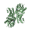
 Assembly


 Mass: 398.437 Da / Num. of mol.: 3 / Source method: obtained synthetically / Formula: C15H22N6O5S
 Type: L-peptide linking / Mass: 146.144 Da / Num. of mol.: 3 / Source method: obtained synthetically / Formula: C5H10N2O3
 Mass: 18.015 Da / Num. of mol.: 236 / Source method: isolated from a natural source / Formula: H2O
 Sample preparation
 / Beamline: X6A / Wavelength: 0.95 Å
 Processing