 Movie
Movie Controller

[English] 日本語
 Yorodumi
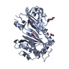
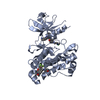
- PDB-1sdj: X-RAY STRUCTURE OF YDDE_ECOLI NORTHEAST STRUCTURAL GENOMICS CONSO... -
+ Open data

- Basic information
| Entry | Database: PDB / ID: 1sdj | |||||||||
|---|---|---|---|---|---|---|---|---|---|---|
| Title | X-RAY STRUCTURE OF YDDE_ECOLI NORTHEAST STRUCTURAL GENOMICS CONSORTIUM TARGET ET25. | |||||||||
 Components Components | Hypothetical protein yddE | |||||||||
 Keywords Keywords | STRUCTURAL GENOMICS / UNKNOWN FUNCTION / PSI / Protein Structure Initiative / Northeast Structural Genomics Consortium / NESG | |||||||||
| Function / homology |  Function and homology information Function and homology informationIsomerases; Racemases and epimerases / isomerase activity / protein homodimerization activity Similarity search - Function | |||||||||
| Biological species |  | |||||||||
| Method |  X-RAY DIFFRACTION / SYNCHROTRON / MAD / Resolution: 2.3 Å X-RAY DIFFRACTION / SYNCHROTRON / MAD / Resolution: 2.3 Å | |||||||||
 Authors Authors | Kuzin, A.P. / Edstrom, W. / Skarina, T. / Korniyenko, Y. / Savchenko, A. / Tong, L. / Northeast Structural Genomics Consortium (NESG) | |||||||||
 Citation Citation | Journal: Proc.Natl.Acad.Sci.Usa / Year: 2004 Title: Structure and function of the phenazine biosynthetic protein PhzF from Pseudomonas fluorescens. Authors: Blankenfeldt, W. / Kuzin, A.P. / Skarina, T. / Korniyenko, Y. / Tong, L. / Bayer, P. / Janning, P. / Thomashow, L.S. / Mavrodi, D.V. | |||||||||
| History |
|
- Structure visualization
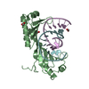
| Structure viewer | Molecule: MolmilJmol/JSmol |
|---|
- Downloads & links
-Download
| PDBx/mmCIF format | 1sdj.cif.gz | 76.5 KB | Display | PDBx/mmCIF format |
|---|---|---|---|---|
| PDB format | pdb1sdj.ent.gz | 56.7 KB | Display | PDB format |
| PDBx/mmJSON format | 1sdj.json.gz | Tree view | PDBx/mmJSON format | |
| Others |  Other downloads Other downloads |
-Validation report
| Arichive directory | https://data.pdbj.org/pub/pdb/validation_reports/sd/1sdjftp://data.pdbj.org/pub/pdb/validation_reports/sd/1sdj | HTTPS FTP |
|---|
-Related structure data
| Related structure data |  1u1vC  1u1wC  1u1xC  1xuaC  1xubC C: citing same article ( |
|---|---|
| Similar structure data | |
| Other databases |


-Links
 PDBj
PDBj- Assembly
| Deposited unit | 
| ||||||||
|---|---|---|---|---|---|---|---|---|---|
| 1 |
| ||||||||
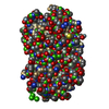
| Unit cell |
|
-Components
| #1: Protein | Mass: 34074.613 Da / Num. of mol.: 1 Source method: isolated from a genetically manipulated source Source: (gene. exp.) | ||||
|---|---|---|---|---|---|
| #2: Chemical | ChemComp-SO4 /   Mass: 96.063 Da / Num. of mol.: 5 / Source method: obtained synthetically / Formula: SO4 Mass: 96.063 Da / Num. of mol.: 5 / Source method: obtained synthetically / Formula: SO4#3: Water | ChemComp-HOH / |  Mass: 18.015 Da / Num. of mol.: 144 / Source method: isolated from a natural source / Formula: H2O Mass: 18.015 Da / Num. of mol.: 144 / Source method: isolated from a natural source / Formula: H2OHas protein modification | Y | |
-Experimental details
-Experiment
| Experiment | Method: X-RAY DIFFRACTION / Number of used crystals: 1 |
|---|
- Sample preparation
| Crystal | Density Matthews: 2.91 Å3/Da / Density % sol: 57.73 % | ||||||||||||||||||||||||||||||||||||||||||
|---|---|---|---|---|---|---|---|---|---|---|---|---|---|---|---|---|---|---|---|---|---|---|---|---|---|---|---|---|---|---|---|---|---|---|---|---|---|---|---|---|---|---|---|
| Crystal grow | *PLUS Method: vapor diffusion, hanging drop / PH range low: 5 / PH range high: 4.4 | ||||||||||||||||||||||||||||||||||||||||||
| Components of the solutions | *PLUS
|
-Data collection
| Diffraction | Mean temperature: 100 K | ||||||||||||
|---|---|---|---|---|---|---|---|---|---|---|---|---|---|
| Diffraction source | Source: SYNCHROTRON / Site: NSLS  / Beamline: X4A / Wavelength: 0.97929, 0.97970, 0.97702 / Beamline: X4A / Wavelength: 0.97929, 0.97970, 0.97702 | ||||||||||||
| Detector | Type: ADSC QUANTUM 4 / Detector: CCD / Date: Apr 11, 2003 | ||||||||||||
| Radiation | Protocol: MAD / Monochromatic (M) / Laue (L): M / Scattering type: x-ray | ||||||||||||
| Radiation wavelength |
| ||||||||||||
| Reflection | Resolution: 2.3→20 Å / Num. all: 140762 / Num. obs: 32871 / % possible obs: 95.7 % / Observed criterion σ(I): -3 / Redundancy: 4.3 % / Biso Wilson estimate: 37.6 Å2 / Rmerge(I) obs: 0.075 / Rsym value: 0.075 | ||||||||||||
| Reflection shell | Resolution: 2.3→2.38 Å / Rmerge(I) obs: 0.247 / Num. unique all: 2943 / Rsym value: 0.247 / % possible all: 85.6 | ||||||||||||
| Reflection | *PLUS % possible obs: 89.5 % | ||||||||||||
| Reflection shell | *PLUS Highest resolution: 2.3 Å / % possible obs: 85.6 % / Redundancy: 27 % / Mean I/σ(I) obs: 3 |
- Processing
| Software |
| ||||||||||||||||||||
|---|---|---|---|---|---|---|---|---|---|---|---|---|---|---|---|---|---|---|---|---|---|
| Refinement | Method to determine structure: MAD / Resolution: 2.3→19.87 Å / Rfactor Rfree error: 0.007 / Data cutoff high absF: 591533.1 / Data cutoff low absF: 0 / Isotropic thermal model: RESTRAINED / Cross valid method: THROUGHOUT / σ(F): 2
| ||||||||||||||||||||
| Solvent computation | Solvent model: FLAT MODEL / Bsol: 38.7607 Å2 / ksol: 0.333235 e/Å3 | ||||||||||||||||||||
| Displacement parameters | Biso mean: 33.7 Å2
| ||||||||||||||||||||
| Refine analyze |
| ||||||||||||||||||||
| Refinement step | Cycle: LAST / Resolution: 2.3→19.87 Å
| ||||||||||||||||||||
| Refine LS restraints |
| ||||||||||||||||||||
| LS refinement shell | Resolution: 2.3→2.44 Å / Rfactor Rfree error: 0.023 / Total num. of bins used: 6
| ||||||||||||||||||||
| Xplor file |
| ||||||||||||||||||||
| Refinement | *PLUS Highest resolution: 2.3 Å / Lowest resolution: 20 Å / % reflection Rfree: 5 % / Rfactor Rfree: 0.26 | ||||||||||||||||||||
| Solvent computation | *PLUS | ||||||||||||||||||||
| Displacement parameters | *PLUS | ||||||||||||||||||||
| Refine LS restraints | *PLUS
|
