 Movie
Movie Controller

[English] 日本語
 Yorodumi
- PDB-1sb8: Crystal structure of Pseudomonas aeruginosa UDP-N-acetylglucosami... -
+ Open data

- Basic information
| Entry | Database: PDB / ID: 1sb8 | ||||||
|---|---|---|---|---|---|---|---|
| Title | Crystal structure of Pseudomonas aeruginosa UDP-N-acetylglucosamine 4-epimerase complexed with UDP-N-acetylgalactosamine | ||||||
 Components Components | wbpP | ||||||
 Keywords Keywords | ISOMERASE / WbpP / epimerase / 4-epimerase / UDP-GalNAc / UDP-GlcNAc / SDR / GalE / NAD / SYK / pseudomonas aeruginosa / UDP / N-acetylglucosamine / N-acetylgalactosamine / UDP-Glc | ||||||
| Function / homology |  Function and homology information Function and homology informationUDP-N-acetyl-alpha-D-glucosaminouronate 4-epimerase / UDP-N-acetylglucosamine 4-epimerase / lipopolysaccharide biosynthetic process / isomerase activity / cell wall organization / nucleotide binding Similarity search - Function | ||||||
| Biological species |   Pseudomonas aeruginosa (bacteria) Pseudomonas aeruginosa (bacteria) | ||||||
| Method |  X-RAY DIFFRACTION / SYNCHROTRON / MOLECULAR REPLACEMENT / Resolution: 2.1 Å X-RAY DIFFRACTION / SYNCHROTRON / MOLECULAR REPLACEMENT / Resolution: 2.1 Å | ||||||
 Authors Authors | Ishiyama, N. / Creuzenet, C. / Lam, J.S. / Berghuis, A.M. | ||||||
 Citation Citation | Journal: J.Biol.Chem. / Year: 2004 Title: Crystal Structure of WbpP, a Genuine UDP-N-acetylglucosamine 4-Epimerase from Pseudomonas aeruginosa: SUBSTRATE SPECIFICITY IN UDP-HEXOSE 4-EPIMERASES. Authors: Ishiyama, N. / Creuzenet, C. / Lam, J.S. / Berghuis, A.M. | ||||||
| History |
|
- Structure visualization
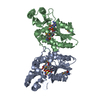
| Structure viewer | Molecule: MolmilJmol/JSmol |
|---|
- Downloads & links
-Download
| PDBx/mmCIF format | 1sb8.cif.gz | 88.5 KB | Display | PDBx/mmCIF format |
|---|---|---|---|---|
| PDB format | pdb1sb8.ent.gz | 64.4 KB | Display | PDB format |
| PDBx/mmJSON format | 1sb8.json.gz | Tree view | PDBx/mmJSON format | |
| Others |  Other downloads Other downloads |
-Validation report
| Summary document | 1sb8_validation.pdf.gz | 1 MB | Display | wwPDB validaton report |
|---|---|---|---|---|
| Full document | 1sb8_full_validation.pdf.gz | 1 MB | Display | |
| Data in XML | 1sb8_validation.xml.gz | 17 KB | Display | |
| Data in CIF | 1sb8_validation.cif.gz | 24.1 KB | Display | |
| Arichive directory | https://data.pdbj.org/pub/pdb/validation_reports/sb/1sb8ftp://data.pdbj.org/pub/pdb/validation_reports/sb/1sb8 | HTTPS FTP |
-Related structure data
| Related structure data | 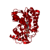 1sb9SC S: Starting model for refinement C: citing same article ( |
|---|---|
| Similar structure data |


-Links
 PDBj
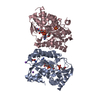
PDBj- Assembly
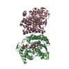
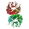
| Deposited unit | 
| ||||||||
|---|---|---|---|---|---|---|---|---|---|
| 1 | 
| ||||||||
| Unit cell |
| ||||||||
| Details | The biological assembly is a homodimer generated from the monomer in the asymmetric unit by the operation: x, -y, -z+1 |
-Components
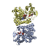
| #1: Protein | Mass: 39080.000 Da / Num. of mol.: 1 Source method: isolated from a genetically manipulated source Source: (gene. exp.) Pseudomonas aeruginosa (bacteria) / Gene: wbpP / Plasmid: pET23 derivative / Production host: References: GenBank: 20560072, UniProt: Q8KN66*PLUS, UDP-N-acetylglucosamine 4-epimerase |
|---|---|
| #2: Chemical | ChemComp-NAD /   Mass: 663.425 Da / Num. of mol.: 1 / Source method: obtained synthetically / Formula: C21H27N7O14P2 / Comment: NAD*YM Mass: 663.425 Da / Num. of mol.: 1 / Source method: obtained synthetically / Formula: C21H27N7O14P2 / Comment: NAD*YM |
| #3: Chemical | ChemComp-UD2 / 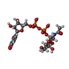  Mass: 607.354 Da / Num. of mol.: 1 / Source method: obtained synthetically / Formula: C17H27N3O17P2 Mass: 607.354 Da / Num. of mol.: 1 / Source method: obtained synthetically / Formula: C17H27N3O17P2 |
| #4: Water | ChemComp-HOH /  Mass: 18.015 Da / Num. of mol.: 182 / Source method: isolated from a natural source / Formula: H2O Mass: 18.015 Da / Num. of mol.: 182 / Source method: isolated from a natural source / Formula: H2O |
-Experimental details
-Experiment
| Experiment | Method: X-RAY DIFFRACTION / Number of used crystals: 1 |
|---|
- Sample preparation
| Crystal | Density Matthews: 2.63 Å3/Da / Density % sol: 52.9 % |
|---|---|
| Crystal grow | Temperature: 295 K / Method: vapor diffusion, hanging drop / pH: 4.5 Details: PEG 600, phosphate-citrate buffer, pH 4.5, VAPOR DIFFUSION, HANGING DROP, temperature 295K |
-Data collection
| Diffraction | Mean temperature: 95 K |
|---|---|
| Diffraction source | Source: SYNCHROTRON / Site: NSLS  / Beamline: X8C / Wavelength: 0.98 / Beamline: X8C / Wavelength: 0.98 |
| Detector | Type: ADSC QUANTUM 4 / Detector: CCD / Date: Nov 6, 2002 |
| Radiation | Protocol: SINGLE WAVELENGTH / Monochromatic (M) / Laue (L): M / Scattering type: x-ray |
| Radiation wavelength | Wavelength: 0.98 Å / Relative weight: 1 |
| Reflection | Resolution: 2.1→41.25 Å / Num. all: 24082 / Num. obs: 23213 / % possible obs: 96.4 % / Observed criterion σ(F): 0 / Observed criterion σ(I): 0 / Redundancy: 6.4 % / Biso Wilson estimate: 11.9 Å2 / Rsym value: 0.083 / Net I/σ(I): 8.7 |
| Reflection shell | Resolution: 2.1→2.18 Å / Redundancy: 6.6 % / Mean I/σ(I) obs: 2.3 / Num. unique all: 2271 / Rsym value: 0.328 / % possible all: 89.7 |
- Processing
| Software |
| ||||||||||||||||||||||||||||||||||||
|---|---|---|---|---|---|---|---|---|---|---|---|---|---|---|---|---|---|---|---|---|---|---|---|---|---|---|---|---|---|---|---|---|---|---|---|---|---|
| Refinement | Method to determine structure: MOLECULAR REPLACEMENT Starting model: partially refined model of PDB entry 1SB9 Resolution: 2.1→41.25 Å / Rfactor Rfree error: 0.005 / Data cutoff high absF: 145620.51 / Data cutoff high rms absF: 145620.51 / Data cutoff low absF: 0 / Isotropic thermal model: RESTRAINED / Cross valid method: THROUGHOUT / σ(F): 0 / Stereochemistry target values: Engh & Huber
| ||||||||||||||||||||||||||||||||||||
| Solvent computation | Solvent model: FLAT MODEL / Bsol: 59.1887 Å2 / ksol: 0.392563 e/Å3 | ||||||||||||||||||||||||||||||||||||
| Displacement parameters | Biso mean: 22.3 Å2
| ||||||||||||||||||||||||||||||||||||
| Refine analyze |
| ||||||||||||||||||||||||||||||||||||
| Refinement step | Cycle: LAST / Resolution: 2.1→41.25 Å
| ||||||||||||||||||||||||||||||||||||
| Refine LS restraints |
| ||||||||||||||||||||||||||||||||||||
| LS refinement shell | Resolution: 2.1→2.23 Å / Rfactor Rfree error: 0.014 / Total num. of bins used: 6
| ||||||||||||||||||||||||||||||||||||
| Xplor file |
|
