 Movie
Movie Controller

[English] 日本語
 Yorodumi
- PDB-1s8d: Structural basis for degenerate recognition of HIV peptide varian... -
+ Open data

- Basic information
| Entry | Database: PDB / ID: 1s8d | ||||||
|---|---|---|---|---|---|---|---|
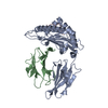
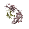
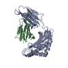
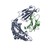
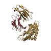
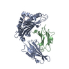
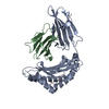
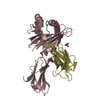
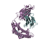
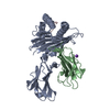
| Title | Structural basis for degenerate recognition of HIV peptide variants by cytotoxic lymphocyte, variant SL9-3A | ||||||
 Components Components |
| ||||||
 Keywords Keywords | IMMUNE SYSTEM / CTL / cytotoxic T lymphocytes / HIV / human immunodeficiency virus / MHC / major histocompatibility complex / pMHC / peptide-MHC complex / RMSD / root-mean-squared deviation / SIV / simian immunodeficiency virus / TCR / T-cell receptor | ||||||
| Function / homology |  Function and homology information Function and homology information: / : / : / : / : / negative regulation of receptor binding / retina homeostasis / positive regulation of protein binding / positive regulation of memory T cell activation / T cell mediated cytotoxicity directed against tumor cell target ...: / : / : / : / : / negative regulation of receptor binding / retina homeostasis / positive regulation of protein binding / positive regulation of memory T cell activation / T cell mediated cytotoxicity directed against tumor cell target / positive regulation of CD8-positive, alpha-beta T cell activation / CD8-positive, alpha-beta T cell activation / positive regulation of CD8-positive, alpha-beta T cell proliferation / antigen processing and presentation of endogenous peptide antigen via MHC class I via ER pathway, TAP-dependent / TAP complex binding / antigen processing and presentation of exogenous peptide antigen via MHC class I / Golgi medial cisterna / regulation of membrane depolarization / CD8 receptor binding / protection from natural killer cell mediated cytotoxicity / beta-2-microglobulin binding / endoplasmic reticulum exit site / TAP binding / detection of bacterium / antigen processing and presentation of endogenous peptide antigen via MHC class Ib / antigen processing and presentation of endogenous peptide antigen via MHC class I via ER pathway, TAP-independent / T cell receptor binding / early endosome lumen / Nef mediated downregulation of MHC class I complex cell surface expression / DAP12 interactions / Endosomal/Vacuolar pathway / T cell mediated cytotoxicity / Antigen Presentation: Folding, assembly and peptide loading of class I MHC / lumenal side of endoplasmic reticulum membrane / regulation of iron ion transport / cellular response to iron(III) ion / negative regulation of iron ion transport / negative regulation of forebrain neuron differentiation / antigen processing and presentation of exogenous protein antigen via MHC class Ib, TAP-dependent / iron ion transport / peptide antigen assembly with MHC class I protein complex / ER to Golgi transport vesicle membrane / regulation of erythrocyte differentiation / response to molecule of bacterial origin / HFE-transferrin receptor complex / MHC class I peptide loading complex / transferrin transport / cellular response to iron ion / negative regulation of receptor-mediated endocytosis / positive regulation of T cell cytokine production / antigen processing and presentation of endogenous peptide antigen via MHC class I / MHC class I protein complex / peptide antigen assembly with MHC class II protein complex / negative regulation of neurogenesis / MHC class II protein complex / cellular response to nicotine / positive regulation of receptor-mediated endocytosis / multicellular organismal-level iron ion homeostasis / positive regulation of T cell mediated cytotoxicity / specific granule lumen / antigen processing and presentation of exogenous peptide antigen via MHC class II / positive regulation of immune response / peptide antigen binding / positive regulation of type II interferon production / phagocytic vesicle membrane / recycling endosome membrane / positive regulation of T cell activation / negative regulation of epithelial cell proliferation / Interferon gamma signaling / Immunoregulatory interactions between a Lymphoid and a non-Lymphoid cell / Interferon alpha/beta signaling / sensory perception of smell / Modulation by Mtb of host immune system / positive regulation of cellular senescence / tertiary granule lumen / MHC class II protein complex binding / T cell differentiation in thymus / DAP12 signaling / late endosome membrane / antimicrobial humoral immune response mediated by antimicrobial peptide / negative regulation of neuron projection development / T cell receptor signaling pathway / antibacterial humoral response / E3 ubiquitin ligases ubiquitinate target proteins / protein refolding / cellular response to lipopolysaccharide / ER-Phagosome pathway / early endosome membrane / amyloid fibril formation / protein homotetramerization / defense response to Gram-negative bacterium / intracellular iron ion homeostasis / learning or memory / defense response to Gram-positive bacterium / immune response / endoplasmic reticulum lumen / Amyloid fiber formation / signaling receptor binding / Golgi membrane / external side of plasma membrane Similarity search - Function | ||||||
| Biological species |  Homo sapiens (human) Homo sapiens (human) | ||||||
| Method |  X-RAY DIFFRACTION / SYNCHROTRON / MOLECULAR REPLACEMENT / Resolution: 2.2 Å X-RAY DIFFRACTION / SYNCHROTRON / MOLECULAR REPLACEMENT / Resolution: 2.2 Å | ||||||
 Authors Authors | Martinez-Hackert, E. / Anikeeva, N. / Kalams, S.A. / Walker, B.D. / Hendrickson, W.A. / Sykulev, Y. | ||||||
 Citation Citation | Journal: J.Biol.Chem. / Year: 2006 Title: Structural Basis for Degenerate Recognition of Natural HIV Peptide Variants by Cytotoxic Lymphocytes. Authors: Martinez-Hackert, E. / Anikeeva, N. / Kalams, S.A. / Walker, B.D. / Hendrickson, W.A. / Sykulev, Y. | ||||||
| History |
|
- Structure visualization
| Structure viewer | Molecule: MolmilJmol/JSmol |
|---|
- Downloads & links
-Download
| PDBx/mmCIF format | 1s8d.cif.gz | 98.7 KB | Display | PDBx/mmCIF format |
|---|---|---|---|---|
| PDB format | pdb1s8d.ent.gz | 74.7 KB | Display | PDB format |
| PDBx/mmJSON format | 1s8d.json.gz | Tree view | PDBx/mmJSON format | |
| Others |  Other downloads Other downloads |
-Validation report
| Arichive directory | https://data.pdbj.org/pub/pdb/validation_reports/s8/1s8dftp://data.pdbj.org/pub/pdb/validation_reports/s8/1s8d | HTTPS FTP |
|---|
-Related structure data
| Related structure data |  1t1wC  1t1xC  1t1yC  1t1zC  1t20C  1t21C  1t22C 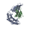 2hlaS S: Starting model for refinement C: citing same article ( |
|---|---|
| Similar structure data |


-Links
 PDBj
PDBj


- Assembly
| Deposited unit | 
| ||||||||
|---|---|---|---|---|---|---|---|---|---|
| 1 |
| ||||||||
| Unit cell |
|
-Components
| #1: Protein | Mass: 31854.203 Da / Num. of mol.: 1 Source method: isolated from a genetically manipulated source Source: (gene. exp.) Homo sapiens (human) / Gene: HLA-A, HLAA / Production host:  |
|---|---|
| #2: Protein | Mass: 11748.160 Da / Num. of mol.: 1 Source method: isolated from a genetically manipulated source Source: (gene. exp.) Homo sapiens (human) / Gene: B2M / Production host: |
| #3: Protein/peptide | Mass: 889.005 Da / Num. of mol.: 1 / Source method: obtained synthetically Details: The peptide was chemically synthesized. The sequence of the peptide is naturally found in human immunodeficiency virus. References: UniProt: O11818*PLUS |
| #4: Water | ChemComp-HOH /  Mass: 18.015 Da / Num. of mol.: 356 / Source method: isolated from a natural source / Formula: H2O Mass: 18.015 Da / Num. of mol.: 356 / Source method: isolated from a natural source / Formula: H2O |
| Has protein modification | Y |
-Experimental details
-Experiment
| Experiment | Method: X-RAY DIFFRACTION / Number of used crystals: 1 |
|---|
- Sample preparation
| Crystal | Density Matthews: 2.61 Å3/Da / Density % sol: 52.95 % |
|---|---|
| Crystal grow | Temperature: 293 K / pH: 6.5 Details: 15% polyethylene glycol 6000, 25mM MES, pH 6.5, VAPOR DIFFUSION, HANGING DROP, temperature 293K, pH 6.50 |
-Data collection
| Diffraction | Mean temperature: 100 K |
|---|---|
| Diffraction source | Source: SYNCHROTRON / Site: NSLS  / Beamline: X4A / Wavelength: 0.97907 / Beamline: X4A / Wavelength: 0.97907 |
| Detector | Type: ADSC QUANTUM 4 / Detector: CCD / Date: Oct 5, 2001 |
| Radiation | Protocol: SINGLE WAVELENGTH / Monochromatic (M) / Laue (L): M / Scattering type: x-ray |
| Radiation wavelength | Wavelength: 0.97907 Å / Relative weight: 1 |
| Reflection | Resolution: 2.2→20 Å / Num. obs: 20625 / Observed criterion σ(F): 0 / Observed criterion σ(I): 0 / Biso Wilson estimate: 17.6 Å2 / Rmerge(I) obs: 0.058 / Rsym value: 0.058 / Net I/σ(I): 4039.3 |
| Reflection shell | Highest resolution: 2.2 Å / Rmerge(I) obs: 0.096 / Mean I/σ(I) obs: 1882 / Rsym value: 0.096 / % possible all: 85.5 |
- Processing
| Software |
| ||||||||||||||||||||||||||||||||||||||||||||||||||||||||||||||||||||||||||||||||
|---|---|---|---|---|---|---|---|---|---|---|---|---|---|---|---|---|---|---|---|---|---|---|---|---|---|---|---|---|---|---|---|---|---|---|---|---|---|---|---|---|---|---|---|---|---|---|---|---|---|---|---|---|---|---|---|---|---|---|---|---|---|---|---|---|---|---|---|---|---|---|---|---|---|---|---|---|---|---|---|---|---|
| Refinement | Method to determine structure: MOLECULAR REPLACEMENT Starting model: PDB ENTRY 2HLA Resolution: 2.2→11.94 Å / Rfactor Rfree error: 0.008 / Data cutoff high absF: 1112275.34 / Data cutoff low absF: 0 / Isotropic thermal model: RESTRAINED / Cross valid method: THROUGHOUT / σ(F): 0
| ||||||||||||||||||||||||||||||||||||||||||||||||||||||||||||||||||||||||||||||||
| Solvent computation | Solvent model: FLAT MODEL / Bsol: 75.9638 Å2 / ksol: 0.466486 e/Å3 | ||||||||||||||||||||||||||||||||||||||||||||||||||||||||||||||||||||||||||||||||
| Displacement parameters | Biso mean: 23.2 Å2
| ||||||||||||||||||||||||||||||||||||||||||||||||||||||||||||||||||||||||||||||||
| Refine analyze |
| ||||||||||||||||||||||||||||||||||||||||||||||||||||||||||||||||||||||||||||||||
| Refinement step | Cycle: LAST / Resolution: 2.2→11.94 Å
| ||||||||||||||||||||||||||||||||||||||||||||||||||||||||||||||||||||||||||||||||
| Refine LS restraints |
| ||||||||||||||||||||||||||||||||||||||||||||||||||||||||||||||||||||||||||||||||
| LS refinement shell | Resolution: 2.2→2.34 Å / Rfactor Rfree error: 0.025 / Total num. of bins used: 6
| ||||||||||||||||||||||||||||||||||||||||||||||||||||||||||||||||||||||||||||||||
| Xplor file |
|
