 Movie
Movie Controller

[English] 日本語
 Yorodumi
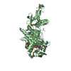
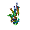
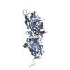
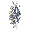
- PDB-1s60: Aminoglycoside N-Acetyltransferase AAC(6')-Iy in Complex with CoA... -
+ Open data

- Basic information
| Entry | Database: PDB / ID: 1s60 | ||||||
|---|---|---|---|---|---|---|---|
| Title | Aminoglycoside N-Acetyltransferase AAC(6')-Iy in Complex with CoA and N-terminal His(6)-tag (crystal form 2) | ||||||
 Components Components | aminoglycoside 6'-N-acetyltransferase | ||||||
 Keywords Keywords | TRANSFERASE / GNAT / N-acetyltransferase / acetyltransferase / aminoglycoside / CoA | ||||||
| Function / homology |  Function and homology information Function and homology informationaminoglycoside 6'-N-acetyltransferase / aminoglycoside 6'-N-acetyltransferase activity / acetyl-CoA metabolic process / response to antibiotic / protein homodimerization activity Similarity search - Function | ||||||
| Biological species |  Salmonella enteritidis (bacteria) Salmonella enteritidis (bacteria) | ||||||
| Method |  X-RAY DIFFRACTION / MOLECULAR REPLACEMENT / Resolution: 3 Å X-RAY DIFFRACTION / MOLECULAR REPLACEMENT / Resolution: 3 Å | ||||||
 Authors Authors | Vetting, M.W. / Magnet, S. / Nieves, E. / Roderick, S.L. / Blanchard, J.S. | ||||||
 Citation Citation | Journal: Chem.Biol. / Year: 2004 Title: A bacterial acetyltransferase capable of regioselective N-acetylation of antibiotics and histones Authors: Vetting, M.W. / Magnet, S. / Nieves, E. / Roderick, S.L. / Blanchard, J.S. #1: Journal: Biochemistry / Year: 2001Title: Kinetic and mutagenic characterization of the chromosomally encoded Salmonella enterica AAC(6')-Iy aminoglycoside N-acetyltransferase Authors: Magnet, S. / Lambert, T. / Courvalin, P. / Blanchard, J. | ||||||
| History |
|
- Structure visualization
| Structure viewer | Molecule: MolmilJmol/JSmol |
|---|
- Downloads & links
-Download
| PDBx/mmCIF format | 1s60.cif.gz | 44.8 KB | Display | PDBx/mmCIF format |
|---|---|---|---|---|
| PDB format | pdb1s60.ent.gz | 31 KB | Display | PDB format |
| PDBx/mmJSON format | 1s60.json.gz | Tree view | PDBx/mmJSON format | |
| Others |  Other downloads Other downloads |
-Validation report
| Arichive directory | https://data.pdbj.org/pub/pdb/validation_reports/s6/1s60ftp://data.pdbj.org/pub/pdb/validation_reports/s6/1s60 | HTTPS FTP |
|---|
-Related structure data


-Links
 PDBj
PDBj


- Assembly
| Deposited unit | 
| ||||||||
|---|---|---|---|---|---|---|---|---|---|
| 1 | 
| ||||||||
| Unit cell |
| ||||||||
| Details | The biological assembly is a dimer generated from the monomer in the assymetric unit by the operations 1 0 0 0 -1 0 0 0 -1 0 146.5 89.0 |
-Components
| #1: Protein | Mass: 18556.926 Da / Num. of mol.: 1 Source method: isolated from a genetically manipulated source Source: (gene. exp.) Salmonella enteritidis (bacteria) / Plasmid: pet28a+ / Production host: References: UniProt: Q9R381, aminoglycoside 6'-N-acetyltransferase |
|---|---|
| #2: Chemical | ChemComp-SO4 /   Mass: 96.063 Da / Num. of mol.: 1 / Source method: obtained synthetically / Formula: SO4 Mass: 96.063 Da / Num. of mol.: 1 / Source method: obtained synthetically / Formula: SO4 |
| #3: Chemical | ChemComp-COA /   Mass: 767.534 Da / Num. of mol.: 1 / Source method: obtained synthetically / Formula: C21H36N7O16P3S Mass: 767.534 Da / Num. of mol.: 1 / Source method: obtained synthetically / Formula: C21H36N7O16P3S |
-Experimental details
-Experiment
| Experiment | Method: X-RAY DIFFRACTION / Number of used crystals: 1 |
|---|
- Sample preparation
| Crystal | Density Matthews: 3.71 Å3/Da / Density % sol: 66.89 % |
|---|---|
| Crystal grow | Temperature: 293 K / Method: vapor diffusion under oil / pH: 8.8 Details: Bicine, Ammonium Sulfate, pH 8.8, vapor diffusion under oil, temperature 293K |
-Data collection
| Diffraction | Mean temperature: 77 K |
|---|---|
| Diffraction source | Source: ROTATING ANODE / Type: RIGAKU RU200 / Wavelength: 1.5418 Å |
| Detector | Type: RIGAKU RAXIS IV / Detector: IMAGE PLATE / Date: Jun 26, 2002 / Details: MSC Blue Confocal |
| Radiation | Monochromator: Optics MSC Blue Confocal / Protocol: SINGLE WAVELENGTH / Monochromatic (M) / Laue (L): M / Scattering type: x-ray |
| Radiation wavelength | Wavelength: 1.5418 Å / Relative weight: 1 |
| Reflection | Resolution: 3→20 Å / Num. all: 5773 / Num. obs: 5773 / % possible obs: 95.3 % / Observed criterion σ(F): 0 / Observed criterion σ(I): 0 / Redundancy: 4 % / Biso Wilson estimate: 71 Å2 / Rsym value: 0.046 / Net I/σ(I): 19.8 |
| Reflection shell | Resolution: 3→3.11 Å / Rsym value: 0.136 / % possible all: 87 |
- Processing
| Software |
| |||||||||||||||||||||||||
|---|---|---|---|---|---|---|---|---|---|---|---|---|---|---|---|---|---|---|---|---|---|---|---|---|---|---|
| Refinement | Method to determine structure: MOLECULAR REPLACEMENT Starting model: Partially refined model created by fitting a Se-MET derived Map from same crystal form collected at synchotron radiation source. Resolution: 3→20 Å / Cross valid method: THROUGHOUT / σ(F): 0 / σ(I): 0 / Stereochemistry target values: Engh & Huber
| |||||||||||||||||||||||||
| Refinement step | Cycle: LAST / Resolution: 3→20 Å
| |||||||||||||||||||||||||
| Refine LS restraints |
| |||||||||||||||||||||||||
| LS refinement shell | Resolution: 3→3.14 Å / Rfactor Rfree: 0.352 / Rfactor Rwork: 0.281 |
