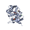
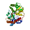
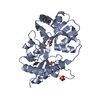
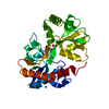
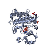
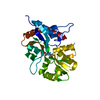
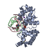
[日本語] English
- PDB-1s50: X-ray structure of the GluR6 ligand binding core (S1S2A) in compl... -

+
データを開く


IDまたはキーワード:

読み込み中...

-
基本情報

登録情報
データベース: PDB / ID: 1s50
タイトルX-ray structure of the GluR6 ligand binding core (S1S2A) in complex with glutamate at 1.65 A resolution
要素Glutamate Receptor 6
キーワードMEMBRANE PROTEIN
機能・相同性
機能・相同性情報


mossy fiber rosette / detection of cold stimulus involved in thermoception / Activation of Na-permeable kainate receptors / kainate selective glutamate receptor complex / Activation of Ca-permeable Kainate Receptor / regulation of short-term neuronal synaptic plasticity / negative regulation of synaptic transmission, glutamatergic / inhibitory postsynaptic potential / glutamate receptor activity / ubiquitin conjugating enzyme binding ...mossy fiber rosette / detection of cold stimulus involved in thermoception / Activation of Na-permeable kainate receptors / kainate selective glutamate receptor complex / Activation of Ca-permeable Kainate Receptor / regulation of short-term neuronal synaptic plasticity / negative regulation of synaptic transmission, glutamatergic / inhibitory postsynaptic potential / glutamate receptor activity / ubiquitin conjugating enzyme binding / regulation of JNK cascade / receptor clustering / modulation of excitatory postsynaptic potential / kainate selective glutamate receptor activity / ionotropic glutamate receptor complex / extracellularly glutamate-gated ion channel activity / behavioral fear response / positive regulation of synaptic transmission / neuronal action potential / glutamate-gated receptor activity / glutamate-gated calcium ion channel activity / ligand-gated monoatomic ion channel activity involved in regulation of presynaptic membrane potential / dendrite cytoplasm / presynaptic modulation of chemical synaptic transmission / hippocampal mossy fiber to CA3 synapse / SNARE binding / regulation of membrane potential / excitatory postsynaptic potential / transmitter-gated monoatomic ion channel activity involved in regulation of postsynaptic membrane potential / synaptic transmission, glutamatergic / PDZ domain binding / modulation of chemical synaptic transmission / regulation of long-term neuronal synaptic plasticity / postsynaptic density membrane / terminal bouton / intracellular calcium ion homeostasis / positive regulation of neuron apoptotic process / presynaptic membrane / scaffold protein binding / neuron apoptotic process / chemical synaptic transmission / perikaryon / negative regulation of neuron apoptotic process / postsynaptic membrane / postsynaptic density / axon / neuronal cell body / synapse / ubiquitin protein ligase binding / dendrite / glutamatergic synapse / identical protein binding / membrane / plasma membrane
類似検索 - 分子機能
Ionotropic glutamate receptor, metazoa / Ligated ion channel L-glutamate- and glycine-binding site / Ionotropic glutamate receptor, L-glutamate and glycine-binding domain / Ligated ion channel L-glutamate- and glycine-binding site / Ligand-gated ion channel / : / Ionotropic glutamate receptor / Eukaryotic homologues of bacterial periplasmic substrate binding proteins. / Receptor, ligand binding region / Receptor family ligand binding region ...Ionotropic glutamate receptor, metazoa / Ligated ion channel L-glutamate- and glycine-binding site / Ionotropic glutamate receptor, L-glutamate and glycine-binding domain / Ligated ion channel L-glutamate- and glycine-binding site / Ligand-gated ion channel / : / Ionotropic glutamate receptor / Eukaryotic homologues of bacterial periplasmic substrate binding proteins. / Receptor, ligand binding region / Receptor family ligand binding region / Periplasmic binding protein-like II / Periplasmic binding protein-like I / D-Maltodextrin-Binding Protein; domain 2 / 3-Layer(aba) Sandwich / Alpha Beta
類似検索 - ドメイン・相同性
GLUTAMIC ACID / Glutamate receptor ionotropic, kainate 2
類似検索 - 構成要素
生物種Rattus norvegicus (ドブネズミ)
手法X線回折 / シンクロトロン / 分子置換 / 解像度: 1.65 Å
データ登録者Mayer, M.L.
引用ジャーナル: Neuron / : 2005
タイトル: Crystal structures of the GluR5 and GluR6 ligand binding cores: Molecular mechanisms underlying kainate receptor selectivity
著者: Mayer, M.L.
履歴
登録2004年1月19日登録サイト: RCSB / 処理サイト: RCSB
改定 1.02005年2月8日Provider: repository / タイプ: Initial release
改定 1.12008年4月29日Group: Version format compliance
改定 1.22011年7月13日Group: Version format compliance
改定 1.32017年7月26日Group: Advisory / Refinement description / Source and taxonomy
カテゴリ: entity_src_gen / pdbx_unobs_or_zero_occ_atoms / software
改定 1.42021年10月27日Group: Advisory / Database references / Derived calculations
カテゴリ: database_2 / pdbx_unobs_or_zero_occ_atoms ...database_2 / pdbx_unobs_or_zero_occ_atoms / struct_ref_seq_dif / struct_site
Item: _database_2.pdbx_DOI / _database_2.pdbx_database_accession ..._database_2.pdbx_DOI / _database_2.pdbx_database_accession / _struct_ref_seq_dif.details / _struct_site.pdbx_auth_asym_id / _struct_site.pdbx_auth_comp_id / _struct_site.pdbx_auth_seq_id
改定 1.52023年8月23日Group: Data collection / Refinement description
カテゴリ: chem_comp_atom / chem_comp_bond / pdbx_initial_refinement_model
改定 1.62024年11月6日Group: Structure summary
カテゴリ: pdbx_entry_details / pdbx_modification_feature
Remark 999 SEQUENCE THE FIRST GLY IS VECTOR ENCODED. THE NATIVE GLUR-5 IS A MEMBRANE PROTEIN. THE PROTEIN ... SEQUENCE THE FIRST GLY IS VECTOR ENCODED. THE NATIVE GLUR-5 IS A MEMBRANE PROTEIN. THE PROTEIN CRYSTALLIZED BY THE AUTHOR IS THE EXTRACELLULAR LIGAND BINDING DOMAIN OF GLUR-5. TRANSMEMBRANE REGIONS WERE GENETICALLY REMOVED AND REPLACED WITH A GLY-THR LINKER. THE SEQUENCE, AS A RESULT, MATCHES DISCONTINUOUSLY WITH THE REFERENCE DATABASE
Remark 300 BIOMOLECULE: 1 THIS ENTRY CONTAINS THE CRYSTALLOGRAPHIC ASYMMETRIC UNIT WHICH CONSISTS OF 1 CHAIN. ... BIOMOLECULE: 1 THIS ENTRY CONTAINS THE CRYSTALLOGRAPHIC ASYMMETRIC UNIT WHICH CONSISTS OF 1 CHAIN. THE BIOLOGICAL UNIT IS BELIEVED TO BE A DIMER, BUT IN THIS CRYSTAL FORM THERE AREN'T ANY SYMMETRY OPERATIONS WHICH GENERATE THE DIMER.

-
構造の表示

構造ビューア分子:
MolmilJmol/JSmol

ダウンロードとリンク

-
集合体

登録構造単位
A: Glutamate Receptor 6
ヘテロ分子


分子量 (理論値)分子数
合計 (水以外)29,5192
ポリマ-29,3721
非ポリマー1471
6,467359
1


  • 登録構造と同一
  • 登録者・ソフトウェアが定義した集合体
タイプ名称対称操作
identity operation1_555x,y,z1
手法PISA
単位格子
Length a, b, c (Å)52.272, 52.272, 169.411
Angle α, β, γ (deg.)90.00, 90.00, 120.00
Int Tables number169
Space group name H-MP61

-
要素

#1: タンパク質 Glutamate Receptor 6


分子量: 29371.633 Da / 分子数: 1 / 断片: GluR6 ligand binding core / 由来タイプ: 組換発現 / 由来: (組換発現) Rattus norvegicus (ドブネズミ) / 遺伝子: GluR6 residues 398-513 and 636-775 / プラスミド: pET22 (modified) / 発現宿主: Escherichia coli (大腸菌) / 株 (発現宿主): Origami B(DE3) / 参照: UniProt: P42260
#2: 化合物 ChemComp-GLU / GLUTAMIC ACID / グルタミン酸


タイプ: L-peptide linking / 分子量: 147.129 Da / 分子数: 1 / 由来タイプ: 合成 / : C5H9NO4
#3: 水 ChemComp-HOH / water


分子量: 18.015 Da / 分子数: 359 / 由来タイプ: 天然 / : H2O
Has protein modificationY

-
実験情報

-
実験

実験手法: X線回折 / 使用した結晶の数: 1

-
試料調製

結晶マシュー密度: 2.31 Å3/Da / 溶媒含有率: 46.31 %
結晶化温度: 293 K / 手法: 蒸気拡散法, ハンギングドロップ法 / pH: 8
詳細: 24% PEG 4000 2 TRIS 20 NaCl 1 EDTA 10 NaGlu , pH 8.0, VAPOR DIFFUSION, HANGING DROP, temperature 293K

-
データ収集

回折平均測定温度: 100 K
放射光源由来: シンクロトロン / サイト: NSLS / ビームライン: X9B / 波長: 0.97946
検出器タイプ: ADSC QUANTUM 4 / 検出器: CCD / 日付: 2003年7月27日
放射モノクロメーター: monochromator / プロトコル: Single Wavelength 0.97946 / 単色(M)・ラウエ(L): M / 散乱光タイプ: x-ray
放射波長波長: 0.97946 Å / 相対比: 1
反射解像度: 1.65→30 Å / Num. all: 31388 / Num. obs: 31253 / % possible obs: 99.6 % / Observed criterion σ(F): 2 / Observed criterion σ(I): 2 / 冗長度: 3.6 % / Biso Wilson estimate: 19.41 Å2 / Rmerge(I) obs: 0.06 / Net I/σ(I): 13.1
反射 シェル解像度: 1.65→1.71 Å / 冗長度: 3.5 % / Rmerge(I) obs: 0.355 / Mean I/σ(I) obs: 3.82 / Num. unique all: 3122 / % possible all: 99.9

-
解析

ソフトウェア
名称分類
HKL-2000データ収集
SCALEPACKデータスケーリング
AMoRE位相決定
CNS精密化
HKL-2000データ削減
精密化構造決定の手法: 分子置換
開始モデル: 1S7Y (rcsb021380)
解像度: 1.65→27.13 Å / Isotropic thermal model: overall anisotropic / 交差検証法: THROUGHOUT / σ(F): 2 / 立体化学のターゲット値: Engh & Huber
Rfactor反射数%反射Selection details
Rfree0.2299 1552 -random
Rwork0.1997 ---
all0.2018 31253 --
obs0.2013 31251 99.4 %-
原子変位パラメータBiso mean: 20.7 Å2
Baniso -1Baniso -2Baniso -3
1-0.84 Å20.84 Å2-1.67 Å2
2--0.89 Å20 Å2
3---0 Å2
Refine analyze
FreeObs
Luzzati coordinate error0.23 Å0.19 Å
Luzzati d res low-5 Å
Luzzati sigma a0.2 Å0.18 Å
精密化ステップサイクル: LAST / 解像度: 1.65→27.13 Å
タンパク質核酸リガンド溶媒全体
原子数2057 0 10 359 2426
拘束条件
Refine-IDタイプDev ideal
X-RAY DIFFRACTIONc_bond_d0.005
X-RAY DIFFRACTIONc_angle_deg1.2
X-RAY DIFFRACTIONc_dihedral_angle_d21.3
X-RAY DIFFRACTIONc_improper_angle_d0.78
LS精密化 シェル解像度: 1.65→1.71 Å / Rfactor Rfree error: 0.018
Rfactor反射数%反射
Rfree0.303 164 -
Rwork0.276 --
obs-3122 99.5 %

+
万見について

-
お知らせ

-
2022年2月9日: EMDBエントリの付随情報ファイルのフォーマットが新しくなりました

EMDBエントリの付随情報ファイルのフォーマットが新しくなりました

  • EMDBのヘッダファイルのバージョン3が、公式のフォーマットとなりました。
  • これまでは公式だったバージョン1.9は、アーカイブから削除されます。

関連情報:EMDBヘッダ

外部リンク:wwPDBはEMDBデータモデルのバージョン3へ移行します

-
2020年8月12日: 新型コロナ情報

新型コロナ情報

URL: https://pdbj.org/emnavi/covid19.php

新ページ: EM Navigatorに新型コロナウイルスの特設ページを開設しました。

関連情報:Covid-19情報 / 2020年3月5日: 新型コロナウイルスの構造データ

+
2020年3月5日: 新型コロナウイルスの構造データ

新型コロナウイルスの構造データ

関連情報:万見生物種 / 2020年8月12日: 新型コロナ情報

外部リンク:COVID-19特集ページ - PDBj / 今月の分子2020年2月:コロナウイルスプロテーアーゼ

+
2019年1月31日: EMDBのIDの桁数の変更

EMDBのIDの桁数の変更

  • EMDBエントリに付与されているアクセスコード(EMDB-ID)は4桁の数字(例、EMD-1234)でしたが、間もなく枯渇します。これまでの4桁のID番号は4桁のまま変更されませんが、4桁の数字を使い切った後に発行されるIDは5桁以上の数字(例、EMD-12345)になります。5桁のIDは2019年の春頃から発行される見通しです。
  • EM Navigator/万見では、接頭語「EMD-」は省略されています。

関連情報:Q: 「EMD」とは何ですか? / 万見/EM NavigatorにおけるID/アクセスコードの表記

外部リンク:EMDB Accession Codes are Changing Soon! / PDBjへお問い合わせ

+
2017年7月12日: PDB大規模アップデート

PDB大規模アップデート

  • 新バージョンのPDBx/mmCIF辞書形式に基づくデータがリリースされました。
  • 今回の更新はバージョン番号が4から5になる大規模なもので、全エントリデータの書き換えが行われる「Remediation」というアップデートに該当します。
  • このバージョンアップで、電子顕微鏡の実験手法に関する多くの項目の書式が改定されました(例:em_softwareなど)。
  • EM NavigatorとYorodumiでも、この改定に基づいた表示内容になります。

外部リンク:wwPDB Remediation / OneDepデータ基準に準拠した、より強化された内容のモデル構造ファイルが、PDBアーカイブで公開されました。

-
万見 (Yorodumi)

幾万の構造データを、幾万の視点から

  • 万見(Yorodumi)は、EMDB/PDB/SASBDBなどの構造データを閲覧するためのページです。
  • EM Navigatorの詳細ページの後継、Omokage検索のフロントエンドも兼ねています。

関連情報:EMDB / PDB / SASBDB / 3つのデータバンクの比較 / 万見検索 / 2016年8月31日: 新しいEM Navigatorと万見 / 万見文献 / Jmol/JSmol / 機能・相同性情報 / 新しいEM Navigatorと万見の変更点

他の情報も見る