 Movie
Movie Controller

[English] 日本語
 Yorodumi
- PDB-1s2u: Crystal structure of the D58A phosphoenolpyruvate mutase mutant p... -
+ Open data

- Basic information
| Entry | Database: PDB / ID: 1s2u | ||||||
|---|---|---|---|---|---|---|---|
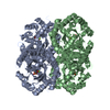
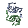
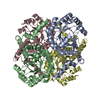
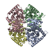
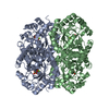
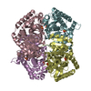
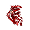
| Title | Crystal structure of the D58A phosphoenolpyruvate mutase mutant protein | ||||||
 Components Components | Phosphoenolpyruvate phosphomutase | ||||||
 Keywords Keywords | ISOMERASE / phosphoenolpyruvate mutase / PEP mutase / phosphonopyruvate / phosphonate biosynthesis pathway | ||||||
| Function / homology |  Function and homology information Function and homology informationphosphoenolpyruvate mutase / phosphoenolpyruvate mutase activity / organic phosphonate biosynthetic process / metal ion binding Similarity search - Function | ||||||
| Biological species |  Mytilus edulis (blue mussel) Mytilus edulis (blue mussel) | ||||||
| Method |  X-RAY DIFFRACTION / SYNCHROTRON / MOLECULAR REPLACEMENT / Resolution: 2 Å X-RAY DIFFRACTION / SYNCHROTRON / MOLECULAR REPLACEMENT / Resolution: 2 Å | ||||||
 Authors Authors | Liu, S. / Lu, Z. / Han, Y. / Jia, Y. / Howard, A. / Dunaway-Mariano, D. / Herzberg, O. | ||||||
 Citation Citation | Journal: Biochemistry / Year: 2004 Title: Conformational Flexibility of PEP Mutase Authors: Liu, S. / Lu, Z. / Han, Y. / Jia, Y. / Howard, A. / Dunaway-Mariano, D. / Herzberg, O. | ||||||
| History |
| ||||||
| Remark 999 | SEQUENCE THE AUTHORS INFORMED THAT THEIR SEQUENCE IS CORRECT AT THE POSITIONS WHERE IT CONFLICTS ...SEQUENCE THE AUTHORS INFORMED THAT THEIR SEQUENCE IS CORRECT AT THE POSITIONS WHERE IT CONFLICTS WITH THE SWISS PROT SEQUENCE. |
- Structure visualization
| Structure viewer | Molecule: MolmilJmol/JSmol |
|---|
- Downloads & links
-Download
| PDBx/mmCIF format | 1s2u.cif.gz | 136.5 KB | Display | PDBx/mmCIF format |
|---|---|---|---|---|
| PDB format | pdb1s2u.ent.gz | 105.7 KB | Display | PDB format |
| PDBx/mmJSON format | 1s2u.json.gz | Tree view | PDBx/mmJSON format | |
| Others |  Other downloads Other downloads |
-Validation report
| Arichive directory | https://data.pdbj.org/pub/pdb/validation_reports/s2/1s2uftp://data.pdbj.org/pub/pdb/validation_reports/s2/1s2u | HTTPS FTP |
|---|
-Related structure data
| Related structure data |  1s2tC  1s2vC  1s2wC  1pymS S: Starting model for refinement C: citing same article ( |
|---|---|
| Similar structure data |


-Links
 PDBj
PDBj
- Assembly
| Deposited unit | 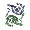
| ||||||||
|---|---|---|---|---|---|---|---|---|---|
| 1 | 
| ||||||||
| Unit cell |
| ||||||||
| Details | biological assembly is a tetramer which can be generated from the dimer by the operation: -x+1, y, -z+1/2 |
-Components
| #1: Protein | Mass: 32910.336 Da / Num. of mol.: 2 / Mutation: D58A Source method: isolated from a genetically manipulated source Source: (gene. exp.) Mytilus edulis (blue mussel) / Plasmid: PET3C / Species (production host): Escherichia coli / Production host:  #2: Chemical |   Mass: 106.120 Da / Num. of mol.: 2 / Source method: obtained synthetically / Formula: C4H10O3 Mass: 106.120 Da / Num. of mol.: 2 / Source method: obtained synthetically / Formula: C4H10O3#3: Water | ChemComp-HOH / |  Mass: 18.015 Da / Num. of mol.: 568 / Source method: isolated from a natural source / Formula: H2O Mass: 18.015 Da / Num. of mol.: 568 / Source method: isolated from a natural source / Formula: H2O |
|---|
-Experimental details
-Experiment
| Experiment | Method: X-RAY DIFFRACTION / Number of used crystals: 1 |
|---|
- Sample preparation
| Crystal | Density Matthews: 2.18 Å3/Da / Density % sol: 43.6 % |
|---|---|
| Crystal grow | Temperature: 298 K / Method: vapor diffusion, hanging drop Details: PEG 4000, Glycerol, HEPES, MgCl2, pH 7.0-8.0, VAPOR DIFFUSION, HANGING DROP, temperature 298K |
-Data collection
| Diffraction | Mean temperature: 100 K |
|---|---|
| Diffraction source | Source: SYNCHROTRON / Site: APS  / Beamline: 17-BM / Wavelength: 1 Å / Beamline: 17-BM / Wavelength: 1 Å |
| Detector | Type: MARRESEARCH / Detector: CCD |
| Radiation | Monochromator: Si(111) double-crystal / Protocol: SINGLE WAVELENGTH / Monochromatic (M) / Laue (L): M / Scattering type: x-ray |
| Radiation wavelength | Wavelength: 1 Å / Relative weight: 1 |
| Reflection | Resolution: 2→50 Å / Num. obs: 36902 / % possible obs: 94.1 % / Observed criterion σ(I): 1 / Redundancy: 5.48 % / Rmerge(I) obs: 0.061 / Net I/σ(I): 10.4 |
| Reflection shell | Resolution: 2→2.05 Å / Rmerge(I) obs: 0.299 / % possible all: 84.5 |
- Processing
| Software |
| |||||||||||||||||||||||||
|---|---|---|---|---|---|---|---|---|---|---|---|---|---|---|---|---|---|---|---|---|---|---|---|---|---|---|
| Refinement | Method to determine structure: MOLECULAR REPLACEMENT Starting model: PDB entry 1pym Resolution: 2→27.7 Å / Isotropic thermal model: isotropic / Cross valid method: THROUGHOUT / σ(F): 2 / Stereochemistry target values: Engh & Huber
| |||||||||||||||||||||||||
| Refinement step | Cycle: LAST / Resolution: 2→27.7 Å
| |||||||||||||||||||||||||
| Refine LS restraints |
|
