 Movie
Movie Controller

[English] 日本語
 Yorodumi
- PDB-1m1b: Crystal Structure of Phosphoenolpyruvate Mutase Complexed with Su... -
+ Open data

- Basic information
| Entry | Database: PDB / ID: 1m1b | ||||||
|---|---|---|---|---|---|---|---|
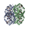
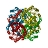
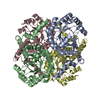
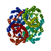
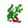
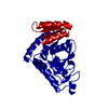
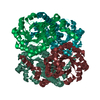
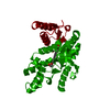
| Title | Crystal Structure of Phosphoenolpyruvate Mutase Complexed with Sulfopyruvate | ||||||
 Components Components | PHOSPHOENOLPYRUVATE PHOSPHOMUTASE | ||||||
 Keywords Keywords | ISOMERASE / phosphoenolpyruvate mutase / PEP mutase / sulfopyruvate | ||||||
| Function / homology |  Function and homology information Function and homology informationphosphoenolpyruvate mutase / phosphoenolpyruvate mutase activity / organic phosphonate biosynthetic process / metal ion binding Similarity search - Function | ||||||
| Biological species |  Mytilus edulis (blue mussel) Mytilus edulis (blue mussel) | ||||||
| Method |  X-RAY DIFFRACTION / MOLECULAR REPLACEMENT / Resolution: 2.25 Å X-RAY DIFFRACTION / MOLECULAR REPLACEMENT / Resolution: 2.25 Å | ||||||
 Authors Authors | Liu, S. / Lu, Z. / Jia, Y. / Dunaway-Mariano, D. / Herzberg, O. | ||||||
 Citation Citation | Journal: Biochemistry / Year: 2002 Title: Dissociative phosphoryl transfer in PEP mutase catalysis: structure of the enzyme/sulfopyruvate complex and kinetic properties of mutants. Authors: Liu, S. / Lu, Z. / Jia, Y. / Dunaway-Mariano, D. / Herzberg, O. | ||||||
| History |
|
- Structure visualization
| Structure viewer | Molecule: MolmilJmol/JSmol |
|---|
- Downloads & links
-Download
| PDBx/mmCIF format | 1m1b.cif.gz | 129.8 KB | Display | PDBx/mmCIF format |
|---|---|---|---|---|
| PDB format | pdb1m1b.ent.gz | 101 KB | Display | PDB format |
| PDBx/mmJSON format | 1m1b.json.gz | Tree view | PDBx/mmJSON format | |
| Others |  Other downloads Other downloads |
-Validation report
| Arichive directory | https://data.pdbj.org/pub/pdb/validation_reports/m1/1m1bftp://data.pdbj.org/pub/pdb/validation_reports/m1/1m1b | HTTPS FTP |
|---|
-Related structure data
| Related structure data |  1pymS S: Starting model for refinement |
|---|---|
| Similar structure data |
-Links
 PDBj
PDBj
- Assembly
| Deposited unit | 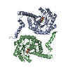
| ||||||||
|---|---|---|---|---|---|---|---|---|---|
| 1 | 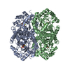
| ||||||||
| Unit cell |
|
-Components
| #1: Protein | Mass: 32954.344 Da / Num. of mol.: 2 Source method: isolated from a genetically manipulated source Source: (gene. exp.) Mytilus edulis (blue mussel) / Plasmid: PET3C / Species (production host): Escherichia coli / Production host:  #2: Chemical |   Mass: 24.305 Da / Num. of mol.: 2 / Source method: obtained synthetically / Formula: Mg Mass: 24.305 Da / Num. of mol.: 2 / Source method: obtained synthetically / Formula: Mg#3: Chemical |   Mass: 168.125 Da / Num. of mol.: 2 / Source method: obtained synthetically / Formula: C3H4O6S Mass: 168.125 Da / Num. of mol.: 2 / Source method: obtained synthetically / Formula: C3H4O6S#4: Water | ChemComp-HOH / |  Mass: 18.015 Da / Num. of mol.: 257 / Source method: isolated from a natural source / Formula: H2O Mass: 18.015 Da / Num. of mol.: 257 / Source method: isolated from a natural source / Formula: H2O |
|---|
-Experimental details
-Experiment
| Experiment | Method: X-RAY DIFFRACTION / Number of used crystals: 1 |
|---|
- Sample preparation
| Crystal | Density Matthews: 2.01 Å3/Da / Density % sol: 38.94 % | |||||||||||||||||||||||||||||||||||||||||||||||||
|---|---|---|---|---|---|---|---|---|---|---|---|---|---|---|---|---|---|---|---|---|---|---|---|---|---|---|---|---|---|---|---|---|---|---|---|---|---|---|---|---|---|---|---|---|---|---|---|---|---|---|
| Crystal grow | Temperature: 298 K / Method: vapor diffusion, hanging drop / pH: 7.5 Details: PEG 4000, glycerol, magnesium chloride, Hepes, sulfopyruvate, pH 7.5, VAPOR DIFFUSION, HANGING DROP, temperature 298K | |||||||||||||||||||||||||||||||||||||||||||||||||
| Crystal grow | *PLUS PH range low: 8 / PH range high: 7 | |||||||||||||||||||||||||||||||||||||||||||||||||
| Components of the solutions | *PLUS
|
-Data collection
| Diffraction | Mean temperature: 100 K |
|---|---|
| Diffraction source | Source: ROTATING ANODE / Type: SIEMENS / Wavelength: 1.5418 Å |
| Detector | Type: MARRESEARCH / Detector: IMAGE PLATE / Date: May 25, 2001 / Details: mirrors |
| Radiation | Monochromator: MIRRORS / Protocol: SINGLE WAVELENGTH / Monochromatic (M) / Laue (L): M / Scattering type: x-ray |
| Radiation wavelength | Wavelength: 1.5418 Å / Relative weight: 1 |
| Reflection | Resolution: 2.25→26.5 Å / Num. all: 25261 / Num. obs: 21045 / % possible obs: 83.3 % / Observed criterion σ(F): 1 / Redundancy: 5.2 % / Rmerge(I) obs: 0.064 / Net I/σ(I): 13.8 |
| Reflection shell | Resolution: 2.25→2.29 Å / Rmerge(I) obs: 0.244 / Mean I/σ(I) obs: 3.7 / % possible all: 64.9 |
| Reflection | *PLUS Rmerge(I) obs: 0.064 |
| Reflection shell | *PLUS % possible obs: 64.9 % / Rmerge(I) obs: 0.244 |
- Processing
| Software |
| |||||||||||||||||||||||||
|---|---|---|---|---|---|---|---|---|---|---|---|---|---|---|---|---|---|---|---|---|---|---|---|---|---|---|
| Refinement | Method to determine structure: MOLECULAR REPLACEMENT Starting model: PDB ENTRY 1PYM Resolution: 2.25→26.5 Å / Cross valid method: THROUGHOUT / σ(F): 2 / Stereochemistry target values: Engh & Huber
| |||||||||||||||||||||||||
| Refinement step | Cycle: LAST / Resolution: 2.25→26.5 Å
| |||||||||||||||||||||||||
| Refine LS restraints |
| |||||||||||||||||||||||||
| Refinement | *PLUS % reflection Rfree: 6 % / Rfactor obs: 0.185 / Rfactor Rfree: 0.268 / Rfactor Rwork: 0.179 | |||||||||||||||||||||||||
| Solvent computation | *PLUS | |||||||||||||||||||||||||
| Displacement parameters | *PLUS | |||||||||||||||||||||||||
| Refine LS restraints | *PLUS Type: c_angle_deg / Dev ideal: 1.5 |
