 Movie
Movie Controller

[English] 日本語
 Yorodumi
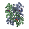
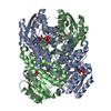
- PDB-1rwb: Cooperative Effect of Two Surface Amino Acid Mutations (Q252L and... -
+ Open data

- Basic information
| Entry | Database: PDB / ID: 1rwb | ||||||
|---|---|---|---|---|---|---|---|
| Title | Cooperative Effect of Two Surface Amino Acid Mutations (Q252L and E170K) of Glucose Dehydrogenase from Bacillus megaterium IWG3 for the stabilization of Oligomeric State | ||||||
 Components Components | Glucose 1-dehydrogenase | ||||||
 Keywords Keywords | OXIDOREDUCTASE | ||||||
| Function / homology |  Function and homology information Function and homology informationglucose 1-dehydrogenase (NAD+) activity / glucose 1-dehydrogenase (NADP+) activity / glucose 1-dehydrogenase [NAD(P)+] / sporulation resulting in formation of a cellular spore Similarity search - Function | ||||||
| Biological species |  Bacillus megaterium (bacteria) Bacillus megaterium (bacteria) | ||||||
| Method |  X-RAY DIFFRACTION / SYNCHROTRON / Rigid Body / Resolution: 2 Å X-RAY DIFFRACTION / SYNCHROTRON / Rigid Body / Resolution: 2 Å | ||||||
 Authors Authors | Baik, S.-H. / Michel, F. / Haser, R. / Harayama, S. | ||||||
 Citation Citation | Journal: Appl.Environ.Microbiol. / Year: 2005 Title: Cooperative effect of two surface amino acid mutations (Q252L and E170K) in glucose dehydrogenase from Bacillus megaterium IWG3 on stabilization of its oligomeric state. Authors: Baik, S.H. / Michel, F. / Aghajari, N. / Haser, R. / Harayama, S. | ||||||
| History |
|
- Structure visualization
| Structure viewer | Molecule: MolmilJmol/JSmol |
|---|
- Downloads & links
-Download
| PDBx/mmCIF format | 1rwb.cif.gz | 216.9 KB | Display | PDBx/mmCIF format |
|---|---|---|---|---|
| PDB format | pdb1rwb.ent.gz | 174.3 KB | Display | PDB format |
| PDBx/mmJSON format | 1rwb.json.gz | Tree view | PDBx/mmJSON format | |
| Others |  Other downloads Other downloads |
-Validation report
| Arichive directory | https://data.pdbj.org/pub/pdb/validation_reports/rw/1rwbftp://data.pdbj.org/pub/pdb/validation_reports/rw/1rwb | HTTPS FTP |
|---|
-Related structure data
| Related structure data | 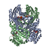 1gcoS S: Starting model for refinement |
|---|---|
| Similar structure data |


-Links
 PDBj
PDBj


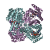
- Assembly
| Deposited unit | 
| ||||||||
|---|---|---|---|---|---|---|---|---|---|
| 1 | 
| ||||||||
| 2 | 
| ||||||||
| Unit cell |
|
-Components
| #1: Protein | Mass: 28098.057 Da / Num. of mol.: 4 / Mutation: Q252L, E170K Source method: isolated from a genetically manipulated source Source: (gene. exp.) Bacillus megaterium (bacteria) / Strain: IWG3 / Production host: References: UniProt: P40288, glucose 1-dehydrogenase [NAD(P)+] #2: Chemical | ChemComp-NAD /   Mass: 663.425 Da / Num. of mol.: 4 / Source method: obtained synthetically / Formula: C21H27N7O14P2 / Comment: NAD*YM Mass: 663.425 Da / Num. of mol.: 4 / Source method: obtained synthetically / Formula: C21H27N7O14P2 / Comment: NAD*YM#3: Water | ChemComp-HOH / |  Mass: 18.015 Da / Num. of mol.: 543 / Source method: isolated from a natural source / Formula: H2O Mass: 18.015 Da / Num. of mol.: 543 / Source method: isolated from a natural source / Formula: H2O |
|---|
-Experimental details
-Experiment
| Experiment | Method: X-RAY DIFFRACTION / Number of used crystals: 1 |
|---|
- Sample preparation
| Crystal | Density Matthews: 2.03 Å3/Da / Density % sol: 39.47 % | ||||||||||||||||||||||||
|---|---|---|---|---|---|---|---|---|---|---|---|---|---|---|---|---|---|---|---|---|---|---|---|---|---|
| Crystal grow | Temperature: 291 K / Method: vapor diffusion, hanging drop / pH: 6.5 Details: PEG 6000, MES, pH 6.5, VAPOR DIFFUSION, HANGING DROP, temperature 291K | ||||||||||||||||||||||||
| Crystal grow | *PLUS pH: 6.3 / Method: vapor diffusion, hanging drop | ||||||||||||||||||||||||
| Components of the solutions | *PLUS
|
-Data collection
| Diffraction | Mean temperature: 100 K |
|---|---|
| Diffraction source | Source: SYNCHROTRON / Site: ESRF  / Beamline: BM30A / Wavelength: 0.95 Å / Beamline: BM30A / Wavelength: 0.95 Å |
| Detector | Type: MARRESEARCH / Detector: CCD / Date: Oct 5, 2002 |
| Radiation | Monochromator: two crystal monochromator between two cylindrical parabolic mirrors Protocol: SINGLE WAVELENGTH / Monochromatic (M) / Laue (L): M / Scattering type: x-ray |
| Radiation wavelength | Wavelength: 0.95 Å / Relative weight: 1 |
| Reflection | Resolution: 1.83→40 Å / Num. obs: 78937 / % possible obs: 99.4 % / Observed criterion σ(F): 1 / Observed criterion σ(I): 1 / Redundancy: 2.8 % / Biso Wilson estimate: 9.5 Å2 / Rmerge(I) obs: 0.073 / Rsym value: 0.06 / Net I/σ(I): 7 |
| Reflection shell | Resolution: 1.83→1.92 Å / Redundancy: 1.8 % / Rmerge(I) obs: 0.19 / Mean I/σ(I) obs: 3.6 / Num. unique all: 2274 / Rsym value: 0.19 / % possible all: 99.4 |
| Reflection | *PLUS Highest resolution: 2 Å / Lowest resolution: 30 Å / Num. obs: 61998 / Redundancy: 3.1 % / Num. measured all: 193166 / Rmerge(I) obs: 0.057 |
| Reflection shell | *PLUS % possible obs: 99.4 % / Redundancy: 3.1 % / Rmerge(I) obs: 0.119 / Mean I/σ(I) obs: 5.9 |
- Processing
| Software |
| ||||||||||||||||||||||||||||||||||||
|---|---|---|---|---|---|---|---|---|---|---|---|---|---|---|---|---|---|---|---|---|---|---|---|---|---|---|---|---|---|---|---|---|---|---|---|---|---|
| Refinement | Method to determine structure: Rigid Body Starting model: PDB Entry 1GCO Resolution: 2→14.99 Å / Rfactor Rfree error: 0.003 / Data cutoff high absF: 2792389.89 / Data cutoff low absF: 0 / Isotropic thermal model: RESTRAINED / Cross valid method: THROUGHOUT / σ(F): 0 / σ(I): 0
| ||||||||||||||||||||||||||||||||||||
| Solvent computation | Solvent model: FLAT MODEL / Bsol: 20.2644 Å2 / ksol: 0.356288 e/Å3 | ||||||||||||||||||||||||||||||||||||
| Displacement parameters | Biso mean: 19.9 Å2
| ||||||||||||||||||||||||||||||||||||
| Refine analyze |
| ||||||||||||||||||||||||||||||||||||
| Refinement step | Cycle: LAST / Resolution: 2→14.99 Å
| ||||||||||||||||||||||||||||||||||||
| Refine LS restraints |
| ||||||||||||||||||||||||||||||||||||
| LS refinement shell | Resolution: 2→2.12 Å / Rfactor Rfree error: 0.011 / Total num. of bins used: 6
| ||||||||||||||||||||||||||||||||||||
| Xplor file |
| ||||||||||||||||||||||||||||||||||||
| Refinement | *PLUS Highest resolution: 2 Å / Lowest resolution: 15 Å / % reflection Rfree: 10 % | ||||||||||||||||||||||||||||||||||||
| Solvent computation | *PLUS | ||||||||||||||||||||||||||||||||||||
| Displacement parameters | *PLUS | ||||||||||||||||||||||||||||||||||||
| Refine LS restraints | *PLUS
|
