 ムービー
ムービー コントローラー


+ データを開く

- 基本情報
基本情報
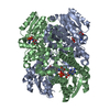
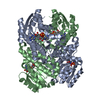
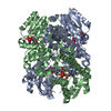
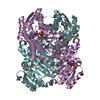
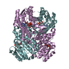
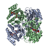
| 登録情報 | データベース: PDB / ID: 1g6k | ||||||
|---|---|---|---|---|---|---|---|
| タイトル | Crystal structure of glucose dehydrogenase mutant E96A complexed with NAD+ | ||||||
 要素 要素 | GLUCOSE 1-DEHYDROGENASE | ||||||
 キーワード キーワード | OXIDOREDUCTASE / short-chain dehydrogenase/reductase | ||||||
| 機能・相同性 |  機能・相同性情報 機能・相同性情報glucose 1-dehydrogenase (NAD+) activity / glucose 1-dehydrogenase (NADP+) activity / glucose 1-dehydrogenase [NAD(P)+] / sporulation resulting in formation of a cellular spore 類似検索 - 分子機能 | ||||||
| 生物種 |  Bacillus megaterium (バクテリア) Bacillus megaterium (バクテリア) | ||||||
| 手法 |  X線回折 / シンクロトロン / 分子置換 / 解像度: 2 Å X線回折 / シンクロトロン / 分子置換 / 解像度: 2 Å | ||||||
 データ登録者 データ登録者 | Yamamoto, K. / Kurisu, G. / Kusunoki, M. / Tabata, S. / Urabe, I. / Osaki, S. | ||||||
 引用 引用 | ジャーナル: To be Published タイトル: Structural analysis of stability-increasing mutants of glucose dehydrogenase 著者: Yamamoto, K. / Kurisu, G. / Kusunoki, M. / Tabata, S. / Urabe, I. / Osaki, S. #1: ジャーナル: J.BIOCHEM.(TOKYO) / 年: 2001タイトル: Crystal structure of glucose dehydrogenase from Bacillus megaterium IWG3 at 1.7 A resolution 著者: Yamamoto, K. / Kurisu, G. / Kusunoki, M. / Tabata, S. / Urabe, I. / Osaki, S. #2: ジャーナル: Acta Crystallogr.,Sect.D / 年: 2000タイトル: Crystallization and preliminary X-ray analysis of glucose dehydrogenase from Bacillus megaterium IWG3 著者: Yamaoto, K. / Kusunoki, M. / Urabe, I. / Tabata, S. / Osaki, S. | ||||||
| 履歴 |
|
- 構造の表示
構造の表示
| 構造ビューア | 分子: MolmilJmol/JSmol |
|---|
- ダウンロードとリンク
-ダウンロード
| PDBx/mmCIF形式 | 1g6k.cif.gz | 210.8 KB | 表示 | PDBx/mmCIF形式 |
|---|---|---|---|---|
| PDB形式 | pdb1g6k.ent.gz | 170.3 KB | 表示 | PDB形式 |
| PDBx/mmJSON形式 | 1g6k.json.gz | ツリー表示 | PDBx/mmJSON形式 | |
| その他 |  その他のダウンロード その他のダウンロード |
-検証レポート
| アーカイブディレクトリ | https://data.pdbj.org/pub/pdb/validation_reports/g6/1g6kftp://data.pdbj.org/pub/pdb/validation_reports/g6/1g6k | HTTPS FTP |
|---|
-関連構造データ

-リンク
 PDBj
PDBj


- 集合体
集合体
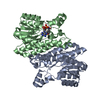
| 登録構造単位 | 
| ||||||||
|---|---|---|---|---|---|---|---|---|---|
| 1 | 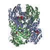
| ||||||||
| 2 | 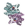
| ||||||||
| 3 | 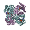
| ||||||||
| 単位格子 |
| ||||||||
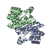
| 詳細 | The biological active form is tetramer. For tetramer1, apply (-x, y, -z)+a to chains A and B. For tetramer2, apply (-x, y, -z)+c to chains E and F. |
-要素
| #1: タンパク質 | 分子量: 28054.924 Da / 分子数: 4 / 変異: E96A / 由来タイプ: 組換発現 由来: (組換発現) Bacillus megaterium (バクテリア)株: IWG3 / プラスミド: PKP1500 / 発現宿主: #2: 化合物 | ChemComp-NAD /   分子量: 663.425 Da / 分子数: 4 / 由来タイプ: 合成 / 式: C21H27N7O14P2 / コメント: NAD*YM 分子量: 663.425 Da / 分子数: 4 / 由来タイプ: 合成 / 式: C21H27N7O14P2 / コメント: NAD*YM#3: 水 | ChemComp-HOH / |  分子量: 18.015 Da / 分子数: 311 / 由来タイプ: 天然 / 式: H2O 分子量: 18.015 Da / 分子数: 311 / 由来タイプ: 天然 / 式: H2O |
|---|
-実験情報
-実験
| 実験 | 手法: X線回折 / 使用した結晶の数: 1 |
|---|
- 試料調製
試料調製
| 結晶 | マシュー密度: 2.08 Å3/Da / 溶媒含有率: 40.42 % |
|---|---|
| 結晶化 | 温度: 293 K / 手法: 蒸気拡散法, ハンギングドロップ法 / pH: 6 詳細: PEG2000, sodium phosphate, pH 6.0, VAPOR DIFFUSION, HANGING DROP, temperature 293K |
-データ収集
| 回折 | 平均測定温度: 288 K |
|---|---|
| 放射光源 | 由来: シンクロトロン / サイト: Photon Factory  / ビームライン: BL-18B / 波長: 1 Å / ビームライン: BL-18B / 波長: 1 Å |
| 検出器 | タイプ: WEISSENBERG / 検出器: DIFFRACTOMETER / 日付: 1998年6月6日 |
| 放射 | プロトコル: SINGLE WAVELENGTH / 単色(M)・ラウエ(L): M / 散乱光タイプ: x-ray |
| 放射波長 | 波長: 1 Å / 相対比: 1 |
| 反射 | 解像度: 2→100 Å / Num. obs: 294512 / % possible obs: 94.6 % / Observed criterion σ(I): 2 / 冗長度: 4.78 % / Biso Wilson estimate: 18.7 Å2 / Rmerge(I) obs: 0.076 / Net I/σ(I): 17.7 |
| 反射 シェル | 解像度: 2→2.07 Å / 冗長度: 2.2 % / Rmerge(I) obs: 0.208 / Num. unique all: 5172 / % possible all: 80.2 |
- 解析
解析
| ソフトウェア |
| |||||||||||||||||||||||||
|---|---|---|---|---|---|---|---|---|---|---|---|---|---|---|---|---|---|---|---|---|---|---|---|---|---|---|
| 精密化 | 構造決定の手法: 分子置換 開始モデル: PDB ENTRY 1GCO 解像度: 2→39.95 Å / 交差検証法: THROUGHOUT / σ(F): 2
| |||||||||||||||||||||||||
| 原子変位パラメータ | Biso mean: 29.9 Å2
| |||||||||||||||||||||||||
| Refine analyze |
| |||||||||||||||||||||||||
| 精密化ステップ | サイクル: LAST / 解像度: 2→39.95 Å
| |||||||||||||||||||||||||
| 拘束条件 |
|
