 Movie
Movie Controller


+ Open data

- Basic information
| Entry | Database: PDB / ID: 1gco | ||||||
|---|---|---|---|---|---|---|---|
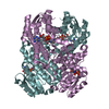
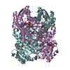
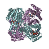
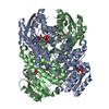
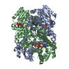
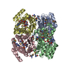
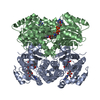
| Title | CRYSTAL STRUCTURE OF GLUCOSE DEHYDROGENASE COMPLEXED WITH NAD+ | ||||||
 Components Components | GLUCOSE DEHYDROGENASE | ||||||
 Keywords Keywords | OXIDOREDUCTASE / short-chain dehydrogenase/reductase | ||||||
| Function / homology |  Function and homology information Function and homology informationglucose 1-dehydrogenase (NAD+) activity / glucose 1-dehydrogenase (NADP+) activity / glucose 1-dehydrogenase [NAD(P)+] / sporulation resulting in formation of a cellular spore Similarity search - Function | ||||||
| Biological species |  Bacillus megaterium (bacteria) Bacillus megaterium (bacteria) | ||||||
| Method |  X-RAY DIFFRACTION / SYNCHROTRON / Resolution: 1.7 Å X-RAY DIFFRACTION / SYNCHROTRON / Resolution: 1.7 Å | ||||||
 Authors Authors | Yamamoto, K. / Kurisu, G. / Kusunoki, M. / Tabata, S. / Urabe, I. / Osaki, S. | ||||||
 Citation Citation | Journal: J.Biochem. / Year: 2001 Title: Crystal structure of glucose dehydrogenase from Bacillus megaterium IWG3 at 1.7 A resolution. Authors: Yamamoto, K. / Kurisu, G. / Kusunoki, M. / Tabata, S. / Urabe, I. / Osaki, S. #1: Journal: Acta Crystallogr.,Sect.D / Year: 2000Title: Crystallization and preliminary X-ray analysis of glucose dehydrogenase from Bacillus megaterium IWG3 Authors: Yamamoto, K. / Kusunoki, M. / Urabe, I. / Tabata, S. / Osaki, S. | ||||||
| History |
|
- Structure visualization
| Structure viewer | Molecule: MolmilJmol/JSmol |
|---|
- Downloads & links
-Download
| PDBx/mmCIF format | 1gco.cif.gz | 216.1 KB | Display | PDBx/mmCIF format |
|---|---|---|---|---|
| PDB format | pdb1gco.ent.gz | 174.5 KB | Display | PDB format |
| PDBx/mmJSON format | 1gco.json.gz | Tree view | PDBx/mmJSON format | |
| Others |  Other downloads Other downloads |
-Validation report
| Arichive directory | https://data.pdbj.org/pub/pdb/validation_reports/gc/1gcoftp://data.pdbj.org/pub/pdb/validation_reports/gc/1gco | HTTPS FTP |
|---|
-Related structure data
| Similar structure data |
|---|


-Links
 PDBj
PDBj


- Assembly
| Deposited unit | 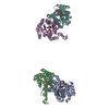
| ||||||||
|---|---|---|---|---|---|---|---|---|---|
| 1 | 
| ||||||||
| 2 | 
| ||||||||
| Unit cell |
|
-Components
| #1: Protein | Mass: 28112.963 Da / Num. of mol.: 4 Source method: isolated from a genetically manipulated source Source: (gene. exp.) Bacillus megaterium (bacteria) / Strain: IWG3 / Plasmid: PKP1500 / Production host: References: UniProt: P40288, glucose 1-dehydrogenase [NAD(P)+] #2: Chemical | ChemComp-NAD /   Mass: 663.425 Da / Num. of mol.: 4 / Source method: obtained synthetically / Formula: C21H27N7O14P2 / Comment: NAD*YM Mass: 663.425 Da / Num. of mol.: 4 / Source method: obtained synthetically / Formula: C21H27N7O14P2 / Comment: NAD*YM#3: Water | ChemComp-HOH / |  Mass: 18.015 Da / Num. of mol.: 562 / Source method: isolated from a natural source / Formula: H2O Mass: 18.015 Da / Num. of mol.: 562 / Source method: isolated from a natural source / Formula: H2O |
|---|
-Experimental details
-Experiment
| Experiment | Method: X-RAY DIFFRACTION / Number of used crystals: 1 |
|---|
- Sample preparation
| Crystal | Density Matthews: 2.13 Å3/Da / Density % sol: 42.26 % | ||||||||||||||||||||||||||||||
|---|---|---|---|---|---|---|---|---|---|---|---|---|---|---|---|---|---|---|---|---|---|---|---|---|---|---|---|---|---|---|---|
| Crystal grow | Temperature: 293 K / Method: vapor diffusion, hanging drop / pH: 6 Details: PEG2000, sodium phosphate, pH 6.0, VAPOR DIFFUSION, HANGING DROP, temperature 293K | ||||||||||||||||||||||||||||||
| Crystal grow | *PLUS Temperature: 294 KDetails: used microseeding, Yamamoto, K., (2000) Acta Crystallogr., Sect.D, 56, 1443. | ||||||||||||||||||||||||||||||
| Components of the solutions | *PLUS
|
-Data collection
| Diffraction | Mean temperature: 290 K |
|---|---|
| Diffraction source | Source: SYNCHROTRON / Site: Photon Factory  / Beamline: BL-18B / Wavelength: 1 / Beamline: BL-18B / Wavelength: 1 |
| Detector | Type: WEISSENBERG / Detector: DIFFRACTOMETER / Date: Jun 18, 1999 |
| Radiation | Protocol: SINGLE WAVELENGTH / Monochromatic (M) / Laue (L): M / Scattering type: x-ray |
| Radiation wavelength | Wavelength: 1 Å / Relative weight: 1 |
| Reflection | Resolution: 1.7→100 Å / Num. obs: 357354 / % possible obs: 91.9 % / Observed criterion σ(I): 2 / Redundancy: 3.77 % / Biso Wilson estimate: 18.7 Å2 / Rmerge(I) obs: 0.033 / Net I/σ(I): 29.2 |
| Reflection shell | Resolution: 1.7→1.76 Å / Redundancy: 3.71 % / Rmerge(I) obs: 0.166 / Num. unique all: 8013 / % possible all: 77.3 |
| Reflection | *PLUS Num. obs: 94821 / Num. measured all: 357354 |
| Reflection shell | *PLUS % possible obs: 77.3 % / Mean I/σ(I) obs: 11.2 |
- Processing
| Software |
| ||||||||||||||||||||||||||||||||||||||||
|---|---|---|---|---|---|---|---|---|---|---|---|---|---|---|---|---|---|---|---|---|---|---|---|---|---|---|---|---|---|---|---|---|---|---|---|---|---|---|---|---|---|
| Refinement | Resolution: 1.7→34.39 Å / Rfactor Rfree error: 0.003 / Data cutoff high absF: 2894287.86 / Data cutoff low absF: 0 / Isotropic thermal model: RESTRAINED / Cross valid method: THROUGHOUT / σ(F): 2
| ||||||||||||||||||||||||||||||||||||||||
| Solvent computation | Solvent model: FLAT MODEL / Bsol: 40.5391 Å2 / ksol: 0.314666 e/Å3 | ||||||||||||||||||||||||||||||||||||||||
| Refine analyze |
| ||||||||||||||||||||||||||||||||||||||||
| Refinement step | Cycle: LAST / Resolution: 1.7→34.39 Å
| ||||||||||||||||||||||||||||||||||||||||
| Refine LS restraints |
| ||||||||||||||||||||||||||||||||||||||||
| LS refinement shell | Resolution: 1.7→1.81 Å / Rfactor Rfree error: 0.009 / Total num. of bins used: 6
| ||||||||||||||||||||||||||||||||||||||||
| Software | *PLUS Name: CNS / Version: 0.9 / Classification: refinement | ||||||||||||||||||||||||||||||||||||||||
| Refinement | *PLUS σ(F): 2 / % reflection Rfree: 5 % | ||||||||||||||||||||||||||||||||||||||||
| Solvent computation | *PLUS | ||||||||||||||||||||||||||||||||||||||||
| Displacement parameters | *PLUS | ||||||||||||||||||||||||||||||||||||||||
| Refine LS restraints | *PLUS
| ||||||||||||||||||||||||||||||||||||||||
| LS refinement shell | *PLUS Rfactor Rfree: 0.241 / % reflection Rfree: 5 % / Rfactor Rwork: 0.216 |
