 Movie
Movie Controller

[English] 日本語
 Yorodumi
- PDB-1ru9: Crystal Structure (A) of u.v.-irradiated cationic cyclization ant... -
+ Open data

- Basic information
| Entry | Database: PDB / ID: 1ru9 | ||||||
|---|---|---|---|---|---|---|---|
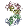
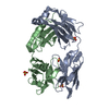
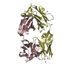
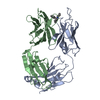
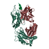
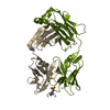
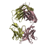
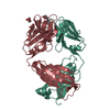
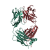
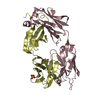
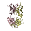
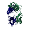
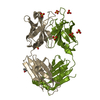
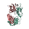
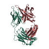
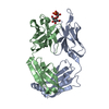
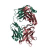
| Title | Crystal Structure (A) of u.v.-irradiated cationic cyclization antibody 4C6 Fab at pH 4.6 with a data set collected in-house. | ||||||
 Components Components | (immunoglobulin igg2a, ...) x 2 | ||||||
 Keywords Keywords | IMMUNE SYSTEM / immunoglobulin / catalytic antibody / water oxidation / amino acid modification | ||||||
| Function / homology |  Function and homology information Function and homology informationpositive regulation of B cell activation / phagocytosis, recognition / humoral immune response mediated by circulating immunoglobulin / early endosome to late endosome transport / positive regulation of type IIa hypersensitivity / positive regulation of type I hypersensitivity / antibody-dependent cellular cytotoxicity / Fc-gamma receptor I complex binding / immunoglobulin complex, circulating / phagocytosis, engulfment ...positive regulation of B cell activation / phagocytosis, recognition / humoral immune response mediated by circulating immunoglobulin / early endosome to late endosome transport / positive regulation of type IIa hypersensitivity / positive regulation of type I hypersensitivity / antibody-dependent cellular cytotoxicity / Fc-gamma receptor I complex binding / immunoglobulin complex, circulating / phagocytosis, engulfment / immunoglobulin receptor binding / IgG immunoglobulin complex / antigen processing and presentation / endosome to lysosome transport / immunoglobulin mediated immune response / regulation of proteolysis / complement activation, classical pathway / positive regulation of endocytosis / antigen binding / multivesicular body / positive regulation of phagocytosis / response to bacterium / positive regulation of immune response / antibacterial humoral response / : / plasma membrane Similarity search - Function | ||||||
| Biological species |  | ||||||
| Method |  X-RAY DIFFRACTION / MOLECULAR REPLACEMENT / Resolution: 2.5 Å X-RAY DIFFRACTION / MOLECULAR REPLACEMENT / Resolution: 2.5 Å | ||||||
 Authors Authors | Zhu, X. / Wentworth Jr., P. / Wentworth, A.D. / Eschenmoser, A. / Lerner, R.A. / Wilson, I.A. | ||||||
 Citation Citation | Journal: Proc.Natl.Acad.Sci.USA / Year: 2004 Title: Probing the antibody-catalyzed water-oxidation pathway at atomic resolution. Authors: Zhu, X. / Wentworth Jr., P. / Wentworth, A.D. / Eschenmoser, A. / Lerner, R.A. / Wilson, I.A. #1: Journal: J.Mol.Biol. / Year: 2003Title: Structural basis for antibody catalysis of a cationic cyclization reaction Authors: Zhu, X. / Heine, A. / Monnat, F. / Houk, K.N. / Janda, K.D. / Wilson, I.A. | ||||||
| History |
| ||||||
| Remark 999 | sequence no suitable sequence data base reference was available at the time of processing this file. |
- Structure visualization
| Structure viewer | Molecule: MolmilJmol/JSmol |
|---|
- Downloads & links
-Download
| PDBx/mmCIF format | 1ru9.cif.gz | 105.6 KB | Display | PDBx/mmCIF format |
|---|---|---|---|---|
| PDB format | pdb1ru9.ent.gz | 79.4 KB | Display | PDB format |
| PDBx/mmJSON format | 1ru9.json.gz | Tree view | PDBx/mmJSON format | |
| Others |  Other downloads Other downloads |
-Validation report
| Arichive directory | https://data.pdbj.org/pub/pdb/validation_reports/ru/1ru9ftp://data.pdbj.org/pub/pdb/validation_reports/ru/1ru9 | HTTPS FTP |
|---|
-Related structure data
| Related structure data | 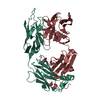 1ruaC  1rukC 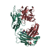 1rulC 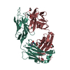 1rumC 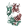 1rupC 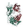 1ruqC 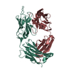 1rurC 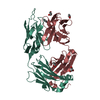 1ncwS S: Starting model for refinement C: citing same article ( |
|---|---|
| Similar structure data |


-Links
 PDBj
PDBj


- Assembly
| Deposited unit | 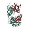
| ||||||||
|---|---|---|---|---|---|---|---|---|---|
| 1 |
| ||||||||
| Unit cell |
|
-Components
-Antibody , 2 types, 2 molecules LH
| #1: Antibody | Mass: 24235.010 Da / Num. of mol.: 1 / Fragment: fab / Source method: isolated from a natural source / Source: (natural) |
|---|---|
| #2: Antibody | Mass: 23832.590 Da / Num. of mol.: 1 / Fragment: fab / Source method: isolated from a natural source / Source: (natural) |
-Non-polymers , 4 types, 249 molecules 


| #3: Chemical | ChemComp-GOL /  Mass: 92.094 Da / Num. of mol.: 5 / Source method: obtained synthetically / Formula: C3H8O3 Mass: 92.094 Da / Num. of mol.: 5 / Source method: obtained synthetically / Formula: C3H8O3#4: Chemical | ChemComp-ACT / |  Mass: 59.044 Da / Num. of mol.: 1 / Source method: obtained synthetically / Formula: C2H3O2 Mass: 59.044 Da / Num. of mol.: 1 / Source method: obtained synthetically / Formula: C2H3O2#5: Chemical | ChemComp-BEZ / |  Mass: 122.121 Da / Num. of mol.: 1 / Source method: obtained synthetically / Formula: C7H6O2 Mass: 122.121 Da / Num. of mol.: 1 / Source method: obtained synthetically / Formula: C7H6O2#6: Water | ChemComp-HOH / | Mass: 18.015 Da / Num. of mol.: 242 / Source method: isolated from a natural source / Formula: H2O |
|---|
-Experimental details
-Experiment
| Experiment | Method: X-RAY DIFFRACTION / Number of used crystals: 1 |
|---|
- Sample preparation
| Crystal | Density Matthews: 2.85 Å3/Da / Density % sol: 56.5 % | ||||||||||||||||||||||||||||||||||||
|---|---|---|---|---|---|---|---|---|---|---|---|---|---|---|---|---|---|---|---|---|---|---|---|---|---|---|---|---|---|---|---|---|---|---|---|---|---|
| Crystal grow | Temperature: 295 K / Method: vapor diffusion, sitting drop / pH: 4.6 Details: 0.1M sodium acetate, pH 4.6, 0.2 M ammonium acetate, 15% (w/v) PEG 4000, VAPOR DIFFUSION, SITTING DROP, temperature 295K | ||||||||||||||||||||||||||||||||||||
| Crystal grow | *PLUS Temperature: 295 K / pH: 5.5 / Method: vapor diffusion, sitting drop | ||||||||||||||||||||||||||||||||||||
| Components of the solutions | *PLUS
|
-Data collection
| Diffraction | Mean temperature: 100 K |
|---|---|
| Diffraction source | Source: ROTATING ANODE / Type: RIGAKU FR-D / Wavelength: 1.5418 Å |
| Detector | Type: MARRESEARCH / Detector: IMAGE PLATE / Date: Mar 28, 2002 |
| Radiation | Protocol: SINGLE WAVELENGTH / Monochromatic (M) / Laue (L): M / Scattering type: x-ray |
| Radiation wavelength | Wavelength: 1.5418 Å / Relative weight: 1 |
| Reflection | Resolution: 2.5→50 Å / Num. obs: 18691 / % possible obs: 92 % / Observed criterion σ(I): -3 / Redundancy: 3.6 % / Rsym value: 0.091 / Net I/σ(I): 12.3 |
| Reflection shell | Resolution: 2.5→2.54 Å / Mean I/σ(I) obs: 2 / Rsym value: 0.447 / % possible all: 90 |
| Reflection | *PLUS Rmerge(I) obs: 0.091 |
| Reflection shell | *PLUS % possible obs: 90.9 % |
- Processing
| Software |
| ||||||||||||||||||||
|---|---|---|---|---|---|---|---|---|---|---|---|---|---|---|---|---|---|---|---|---|---|
| Refinement | Method to determine structure: MOLECULAR REPLACEMENT Starting model: PDB ENTRY 1NCW Resolution: 2.5→50 Å / Cross valid method: FREE R / σ(F): 0 / Stereochemistry target values: Engh & Huber
| ||||||||||||||||||||
| Refinement step | Cycle: LAST / Resolution: 2.5→50 Å
| ||||||||||||||||||||
| Refine LS restraints |
| ||||||||||||||||||||
| Refinement | *PLUS % reflection Rfree: 5 % | ||||||||||||||||||||
| Solvent computation | *PLUS | ||||||||||||||||||||
| Displacement parameters | *PLUS | ||||||||||||||||||||
| Refine LS restraints | *PLUS
|
