 Movie
Movie Controller

[English] 日本語
 Yorodumi
- PDB-1rty: Crystal Structure of Bacillus subtilis YvqK, a putative ATP-bindi... -
+ Open data

- Basic information
| Entry | Database: PDB / ID: 1rty | ||||||
|---|---|---|---|---|---|---|---|
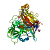
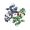
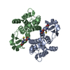
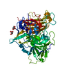
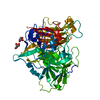
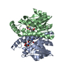
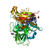
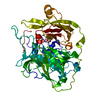
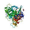
| Title | Crystal Structure of Bacillus subtilis YvqK, a putative ATP-binding Cobalamin Adenosyltransferase, The North East Structural Genomics Target SR128 | ||||||
 Components Components | yvqk protein | ||||||
 Keywords Keywords | STRUCTURAL GENOMICS / UNKNOWN FUNCTION / all alpha-helical trimeric protein / PSI / Protein Structure Initiative / Northeast Structural Genomics Consortium / NESG | ||||||
| Function / homology |  Function and homology information Function and homology informationcorrinoid adenosyltransferase / corrinoid adenosyltransferase activity / porphyrin-containing compound biosynthetic process / cobalamin biosynthetic process / ATP binding / cytoplasm Similarity search - Function | ||||||
| Biological species |  | ||||||
| Method |  X-RAY DIFFRACTION / SYNCHROTRON / MOLECULAR REPLACEMENT / Resolution: 2.4 Å X-RAY DIFFRACTION / SYNCHROTRON / MOLECULAR REPLACEMENT / Resolution: 2.4 Å | ||||||
 Authors Authors | Forouhar, F. / Lee, I. / Xiao, R. / Acton, T.B. / Montelione, G.T. / Tong, L. / Northeast Structural Genomics Consortium (NESG) | ||||||
 Citation Citation | Journal: J.STRUCT.FUNCT.GENOM. / Year: 2007 Title: Functional insights from structural genomics. Authors: Forouhar, F. / Kuzin, A. / Seetharaman, J. / Lee, I. / Zhou, W. / Abashidze, M. / Chen, Y. / Yong, W. / Janjua, H. / Fang, Y. / Wang, D. / Cunningham, K. / Xiao, R. / Acton, T.B. / ...Authors: Forouhar, F. / Kuzin, A. / Seetharaman, J. / Lee, I. / Zhou, W. / Abashidze, M. / Chen, Y. / Yong, W. / Janjua, H. / Fang, Y. / Wang, D. / Cunningham, K. / Xiao, R. / Acton, T.B. / Pichersky, E. / Klessig, D.F. / Porter, C.W. / Montelione, G.T. / Tong, L. | ||||||
| History |
|
- Structure visualization
| Structure viewer | Molecule: MolmilJmol/JSmol |
|---|
- Downloads & links
-Download
| PDBx/mmCIF format | 1rty.cif.gz | 109.5 KB | Display | PDBx/mmCIF format |
|---|---|---|---|---|
| PDB format | pdb1rty.ent.gz | 85.2 KB | Display | PDB format |
| PDBx/mmJSON format | 1rty.json.gz | Tree view | PDBx/mmJSON format | |
| Others |  Other downloads Other downloads |
-Validation report
| Arichive directory | https://data.pdbj.org/pub/pdb/validation_reports/rt/1rtyftp://data.pdbj.org/pub/pdb/validation_reports/rt/1rty | HTTPS FTP |
|---|
-Related structure data
| Related structure data | 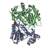 1sqsC 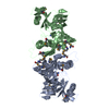 1tm0C 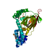 1zbpC 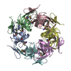 2hd3C 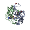 2nv4C 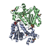 2oysC 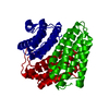 1nogS S: Starting model for refinement C: citing same article ( |
|---|---|
| Similar structure data | |
| Other databases |
-Links
 PDBj
PDBj- Assembly
| Deposited unit | 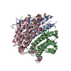
| ||||||||
|---|---|---|---|---|---|---|---|---|---|
| 1 |
| ||||||||
| Unit cell |
| ||||||||
| Details | the protein most likely functions as a trimer. |
-Components
| #1: Protein | Mass: 21613.064 Da / Num. of mol.: 3 Source method: isolated from a genetically manipulated source Source: (gene. exp.) #2: Chemical | ChemComp-PO4 / |   Mass: 94.971 Da / Num. of mol.: 1 / Source method: obtained synthetically / Formula: PO4 Mass: 94.971 Da / Num. of mol.: 1 / Source method: obtained synthetically / Formula: PO4#3: Water | ChemComp-HOH / |  Mass: 18.015 Da / Num. of mol.: 183 / Source method: isolated from a natural source / Formula: H2O Mass: 18.015 Da / Num. of mol.: 183 / Source method: isolated from a natural source / Formula: H2OHas protein modification | Y | |
|---|
-Experimental details
-Experiment
| Experiment | Method: X-RAY DIFFRACTION / Number of used crystals: 1 |
|---|
- Sample preparation
| Crystal | Density Matthews: 2.35 Å3/Da / Density % sol: 47.66 % | |||||||||||||||||||||||||||||||||||||||||||||||||
|---|---|---|---|---|---|---|---|---|---|---|---|---|---|---|---|---|---|---|---|---|---|---|---|---|---|---|---|---|---|---|---|---|---|---|---|---|---|---|---|---|---|---|---|---|---|---|---|---|---|---|
| Crystal grow | Temperature: 277 K / Method: vapor diffusion, hanging drop / pH: 6.2 Details: 50 mM MES, 12% PEG 20,000 , pH 6.2, VAPOR DIFFUSION, HANGING DROP, temperature 277K | |||||||||||||||||||||||||||||||||||||||||||||||||
| Crystal grow | *PLUS pH: 7 / Method: vapor diffusion, hanging drop | |||||||||||||||||||||||||||||||||||||||||||||||||
| Components of the solutions | *PLUS
|
-Data collection
| Diffraction | Mean temperature: 100 K | ||||||||||||
|---|---|---|---|---|---|---|---|---|---|---|---|---|---|
| Diffraction source | Source: SYNCHROTRON / Site: NSLS  / Beamline: X4A / Wavelength: 0.9793, 0.97959, 0.97237 / Beamline: X4A / Wavelength: 0.9793, 0.97959, 0.97237 | ||||||||||||
| Detector | Type: ADSC QUANTUM 4 / Detector: CCD / Date: Jun 15, 2003 | ||||||||||||
| Radiation | Monochromator: monochromator / Protocol: MAD / Monochromatic (M) / Laue (L): M / Scattering type: x-ray | ||||||||||||
| Radiation wavelength |
| ||||||||||||
| Reflection | Resolution: 2.4→24.6 Å / Num. all: 46124 / Num. obs: 39984 / % possible obs: 86.4 % / Observed criterion σ(F): 2 / Observed criterion σ(I): 1 / Redundancy: 4 % / Biso Wilson estimate: 13.6 Å2 / Rmerge(I) obs: 0.101 / Rsym value: 0.108 / Net I/σ(I): 15 | ||||||||||||
| Reflection shell | Resolution: 2.4→2.49 Å / Redundancy: 4 % / Rmerge(I) obs: 0.259 / Mean I/σ(I) obs: 3.99 / Num. unique all: 39984 / Rsym value: 0.283 / % possible all: 86.4 |
- Processing
| Software |
| |||||||||||||||||||||||||
|---|---|---|---|---|---|---|---|---|---|---|---|---|---|---|---|---|---|---|---|---|---|---|---|---|---|---|
| Refinement | Method to determine structure: MOLECULAR REPLACEMENT Starting model: PDB ENTRY 1NOG Resolution: 2.4→24.6 Å / Rfactor Rfree error: 0.005 / Data cutoff high absF: 304817.15 / Data cutoff low absF: 0 / Isotropic thermal model: OVERALL / Cross valid method: THROUGHOUT / σ(F): 2 / σ(I): 2 / Stereochemistry target values: Engh & Huber Details: Only the five N-terminal residues of the monomer B have clear density, as they have been modeled. However, prior to the initiating methionine of the monomer B, there is an additional density ...Details: Only the five N-terminal residues of the monomer B have clear density, as they have been modeled. However, prior to the initiating methionine of the monomer B, there is an additional density of an unrecognizable shape with the molecular mass of 100-150 Dalton. Our mass spectrum also reveals a peak, corresponding to 130 Dalton. Moreover, the unknown chemical entity possibly is covalently bound to the methionine.
| |||||||||||||||||||||||||
| Solvent computation | Solvent model: FLAT MODEL / Bsol: 35.47 Å2 / ksol: 0.320189 e/Å3 | |||||||||||||||||||||||||
| Displacement parameters | Biso mean: 40.9 Å2
| |||||||||||||||||||||||||
| Refine analyze |
| |||||||||||||||||||||||||
| Refinement step | Cycle: LAST / Resolution: 2.4→24.6 Å
| |||||||||||||||||||||||||
| Refine LS restraints |
| |||||||||||||||||||||||||
| LS refinement shell | Resolution: 2.4→2.55 Å / Rfactor Rfree error: 0.013 / Total num. of bins used: 6
| |||||||||||||||||||||||||
| Xplor file |
| |||||||||||||||||||||||||
| Refine LS restraints | *PLUS
|
