 Movie
Movie Controller


+ Open data

- Basic information
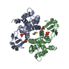
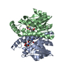
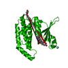
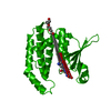
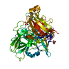
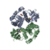
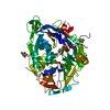
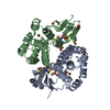
| Entry | Database: PDB / ID: 1oe7 | ||||||
|---|---|---|---|---|---|---|---|
| Title | 28kDa glutathione S-transferase from Schistosoma haematobium | ||||||
 Components Components | GLUTATHIONE S-TRANSFERASE | ||||||
 Keywords Keywords | TRANSFERASE / SCHISTOSOMIASIS / DETOXIFYING ENZYME / PROSTAGLANDIN D2 SYNTHASE / VACCINE CANDIDATE | ||||||
| Function / homology |  Function and homology information Function and homology informationglutathione transferase / glutathione transferase activity / glutathione metabolic process Similarity search - Function | ||||||
| Biological species |  | ||||||
| Method |  X-RAY DIFFRACTION / SYNCHROTRON / MOLECULAR REPLACEMENT / Resolution: 1.8 Å X-RAY DIFFRACTION / SYNCHROTRON / MOLECULAR REPLACEMENT / Resolution: 1.8 Å | ||||||
 Authors Authors | Johnson, K.A. / Angelucci, F. / Tsernoglou, D. | ||||||
 Citation Citation | Journal: Biochemistry / Year: 2003 Title: Crystal Structure of the 28 kDa Glutathione S-Transferase from Schistosoma Haematobium Authors: Johnson, K.A. / Angelucci, F. / Bellelli, A. / Herve, M. / Fontaine, J. / Tsernoglou, D. / Capron, A. / Trottein, F. / Brunori, M. #1: Journal: J.Exp.Med. / Year: 2001 Title: Role of the Parasite-Derived Prostaglandin D2 in the Inhibition of Epidermal Langerhans Cell Migration During Schistosomiasis Infections Authors: Angeli, V. / Faveeuw, C. / Roye, O. / Fontaine, J. / Teissier, E. / Capron, A. / Wolowczuk, L. / Capron, M. / Trottein, F. #2: Journal: J.Mol.Biol. / Year: 1992 Title: Crystallization and Preliminary Diffraction Studies of a Protective Cloned 28 kDa Glutathione S-Transferase from Schistosoma Mansoni Authors: Trottein, F. / Vaney, M.C. / Bachet, B. / Pierce, R.J. / Colloc'H, N. / Lecocq, J.P. / Capron, A. / Momon, J.P. | ||||||
| History |
|
- Structure visualization
| Structure viewer | Molecule: MolmilJmol/JSmol |
|---|
- Downloads & links
-Download
| PDBx/mmCIF format | 1oe7.cif.gz | 101 KB | Display | PDBx/mmCIF format |
|---|---|---|---|---|
| PDB format | pdb1oe7.ent.gz | 77.3 KB | Display | PDB format |
| PDBx/mmJSON format | 1oe7.json.gz | Tree view | PDBx/mmJSON format | |
| Others |  Other downloads Other downloads |
-Validation report
| Arichive directory | https://data.pdbj.org/pub/pdb/validation_reports/oe/1oe7ftp://data.pdbj.org/pub/pdb/validation_reports/oe/1oe7 | HTTPS FTP |
|---|
-Related structure data
| Related structure data | 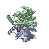 1oe8C 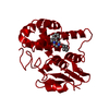 1gtbS C: citing same article ( S: Starting model for refinement |
|---|---|
| Similar structure data |


-Links
 PDBj
PDBj

- Assembly
| Deposited unit | 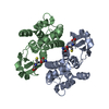
| ||||||||
|---|---|---|---|---|---|---|---|---|---|
| 1 |
| ||||||||
| Unit cell |
| ||||||||
| Noncrystallographic symmetry (NCS) | NCS oper: (Code: given Matrix: (-0.0737, -0.0003, 0.9973), Vector: |
-Components
| #1: Protein | Mass: 23936.746 Da / Num. of mol.: 2 Source method: isolated from a genetically manipulated source Source: (gene. exp.)  #2: Chemical |   Mass: 307.323 Da / Num. of mol.: 2 / Source method: obtained synthetically / Formula: C10H17N3O6S Mass: 307.323 Da / Num. of mol.: 2 / Source method: obtained synthetically / Formula: C10H17N3O6S#3: Water | ChemComp-HOH / |  Mass: 18.015 Da / Num. of mol.: 225 / Source method: isolated from a natural source / Formula: H2O Mass: 18.015 Da / Num. of mol.: 225 / Source method: isolated from a natural source / Formula: H2OSequence details | SEQUENCE MAPPED AGAINST ITSELF PENDING SEQUENCE SUBMISSION TO SWISSPROT DATABASE FOR SCHISTOSOMA ...SEQUENCE MAPPED AGAINST ITSELF PENDING SEQUENCE SUBMISSION | |
|---|
-Experimental details
-Experiment
| Experiment | Method: X-RAY DIFFRACTION / Number of used crystals: 1 |
|---|
- Sample preparation
| Crystal | Density Matthews: 2.3 Å3/Da / Density % sol: 45 % | ||||||||||||||||||||||||||||||
|---|---|---|---|---|---|---|---|---|---|---|---|---|---|---|---|---|---|---|---|---|---|---|---|---|---|---|---|---|---|---|---|
| Crystal grow | Method: vapor diffusion, hanging drop / pH: 8 Details: PROTEIN (~60MG/ML) WAS CRYSTALLIZED IN HANGING DROPS USING A WELL SOLUTION OF 2.1M AMMONIUM SULFATE, 100MM TRIS, PH7.2, 5MM BETA-MERCAPTOETHANOL, pH 8.00 | ||||||||||||||||||||||||||||||
| Crystal grow | *PLUS pH: 7.2 / Method: vapor diffusion, hanging drop | ||||||||||||||||||||||||||||||
| Components of the solutions | *PLUS
|
-Data collection
| Diffraction | Mean temperature: 100 K |
|---|---|
| Diffraction source | Source: SYNCHROTRON / Site: ELETTRA  / Beamline: 5.2R / Wavelength: 1.0032 / Beamline: 5.2R / Wavelength: 1.0032 |
| Detector | Type: MARRESEARCH / Detector: CCD / Date: Oct 15, 2001 |
| Radiation | Protocol: SINGLE WAVELENGTH / Monochromatic (M) / Laue (L): M / Scattering type: x-ray |
| Radiation wavelength | Wavelength: 1.0032 Å / Relative weight: 1 |
| Reflection | Resolution: 1.81→25 Å / Num. obs: 39246 / % possible obs: 96.3 % / Redundancy: 6.5 % / Biso Wilson estimate: 32.9 Å2 / Rmerge(I) obs: 0.071 / Net I/σ(I): 21.5 |
| Reflection shell | Resolution: 1.81→1.86 Å / Redundancy: 2.6 % / Rmerge(I) obs: 0.22 / Mean I/σ(I) obs: 3.4 / % possible all: 72 |
| Reflection | *PLUS Redundancy: 6.5 % / Rmerge(I) obs: 0.071 |
| Reflection shell | *PLUS % possible obs: 72 % / Redundancy: 2.6 % / Rmerge(I) obs: 0.22 / Mean I/σ(I) obs: 3.4 |
- Processing
| Software |
| ||||||||||||||||||||
|---|---|---|---|---|---|---|---|---|---|---|---|---|---|---|---|---|---|---|---|---|---|
| Refinement | Method to determine structure: MOLECULAR REPLACEMENT Starting model: PDB ENTRY 1GTB Resolution: 1.8→20 Å / SU B: 3.62 / SU ML: 0.11 / Cross valid method: THROUGHOUT / ESU R: 0.17 / ESU R Free: 0.17
| ||||||||||||||||||||
| Refinement step | Cycle: LAST / Resolution: 1.8→20 Å
| ||||||||||||||||||||
| Refinement | *PLUS Num. reflection obs: 39246 / % reflection Rfree: 5 % | ||||||||||||||||||||
| Solvent computation | *PLUS | ||||||||||||||||||||
| Displacement parameters | *PLUS | ||||||||||||||||||||
| Refine LS restraints | *PLUS
|
