 Movie
Movie Controller

[English] 日本語
 Yorodumi
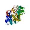
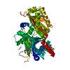
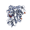
- PDB-1rrv: X-ray crystal structure of TDP-vancosaminyltransferase GtfD as a ... -
+ Open data

- Basic information
| Entry | Database: PDB / ID: 1rrv | |||||||||
|---|---|---|---|---|---|---|---|---|---|---|
| Title | X-ray crystal structure of TDP-vancosaminyltransferase GtfD as a complex with TDP and the natural substrate, desvancosaminyl vancomycin. | |||||||||
 Components Components |
| |||||||||
 Keywords Keywords | TRANSFERASE/ANTIBIOTIC / GT-B / GLYCOSYLTRANSFERASE / ROSSMANN FOLD / GLYCOPEPTIDE / VACOSAMYCIN / ANTIBIOTIC / TRANSFERASE-ANTIBIOTIC COMPLEX | |||||||||
| Function / homology |  Function and homology information Function and homology informationdevancosaminyl-vancomycin vancosaminetransferase / vancomycin biosynthetic process / UDP-glycosyltransferase activity / hexosyltransferase activity / lipid glycosylation / carbohydrate metabolic process Similarity search - Function | |||||||||
| Biological species |  AMYCOLATOPSIS ORIENTALIS (bacteria) AMYCOLATOPSIS ORIENTALIS (bacteria) | |||||||||
| Method |  X-RAY DIFFRACTION / SYNCHROTRON / MOLECULAR REPLACEMENT / Resolution: 2 Å X-RAY DIFFRACTION / SYNCHROTRON / MOLECULAR REPLACEMENT / Resolution: 2 Å | |||||||||
 Authors Authors | Mulichak, A.M. / Lu, W. / Losey, H.C. / Walsh, C.T. / Garavito, R.M. | |||||||||
 Citation Citation | Journal: Biochemistry / Year: 2004 Title: Crystal Structure of Vancosaminyltransferase Gtfd from the Vancomycin Biosynthetic Pathway: Interactions with Acceptor and Nucleotide Ligands Authors: Mulichak, A.M. / Lu, W. / Losey, H.C. / Walsh, C.T. / Garavito, R.M. | |||||||||
| History |
|
- Structure visualization
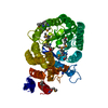
| Structure viewer | Molecule: MolmilJmol/JSmol |
|---|
- Downloads & links
-Download
| PDBx/mmCIF format | 1rrv.cif.gz | 177.8 KB | Display | PDBx/mmCIF format |
|---|---|---|---|---|
| PDB format | pdb1rrv.ent.gz | 137.9 KB | Display | PDB format |
| PDBx/mmJSON format | 1rrv.json.gz | Tree view | PDBx/mmJSON format | |
| Others |  Other downloads Other downloads |
-Validation report
| Arichive directory | https://data.pdbj.org/pub/pdb/validation_reports/rr/1rrvftp://data.pdbj.org/pub/pdb/validation_reports/rr/1rrv | HTTPS FTP |
|---|
-Related structure data
| Related structure data | 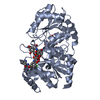 1pn3S S: Starting model for refinement |
|---|---|
| Similar structure data |


-Links
 PDBj
PDBj


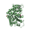
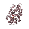
- Assembly
| Deposited unit | 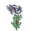
| ||||||||
|---|---|---|---|---|---|---|---|---|---|
| 1 | 
| ||||||||
| 2 | 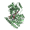
| ||||||||
| Unit cell |
| ||||||||
| Details | Biological assembly is a monomer. |
-Components
-Protein / Protein/peptide / Sugars , 3 types, 6 molecules ABCD
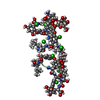
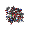
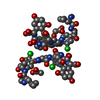
| #1: Protein | Mass: 43905.879 Da / Num. of mol.: 2 Source method: isolated from a genetically manipulated source Source: (gene. exp.) AMYCOLATOPSIS ORIENTALIS (bacteria) / Gene: GTFD / Plasmid: PET22B / Production host: #2: Protein/peptide | 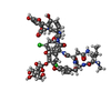  Type: Glycopeptide / Class: Antibiotic / Mass: 1149.977 Da / Num. of mol.: 2 / Source method: isolated from a natural source Type: Glycopeptide / Class: Antibiotic / Mass: 1149.977 Da / Num. of mol.: 2 / Source method: isolated from a natural sourceDetails: DESVANCOSAMINYL VANCOMYCIN IS AN INTERMIDIATE IN VANCOMYCIN BIOSYNTHESIS. IT IS TRICYCLIC GLYCOPEPTIDE, GLYCOSYLATED BY D-GLUCOSE (RESIDUE 8) ON RESIDUE 4. Source: (natural) AMYCOLATOPSIS ORIENTALIS (bacteria) / References: NOR: NOR00681, DESVANCOSAMINYL VANCOMYCIN#6: Sugar |  Type: D-saccharide, beta linking, Glycopeptide / Class: Antibiotic / Mass: 180.156 Da / Num. of mol.: 2 Type: D-saccharide, beta linking, Glycopeptide / Class: Antibiotic / Mass: 180.156 Da / Num. of mol.: 2Source method: isolated from a genetically manipulated source Formula: C6H12O6 Details: DESVANCOSAMINYL VANCOMYCIN IS AN INTERMIDIATE IN VANCOMYCIN BIOSYNTHESIS. IT IS TRICYCLIC GLYCOPEPTIDE, GLYCOSYLATED BY D-GLUCOSE (RESIDUE 8) ON RESIDUE 4. References: DESVANCOSAMINYL VANCOMYCIN |
|---|
-Non-polymers , 4 types, 507 molecules 


| #3: Chemical |  Mass: 39.098 Da / Num. of mol.: 2 / Source method: obtained synthetically / Formula: K Mass: 39.098 Da / Num. of mol.: 2 / Source method: obtained synthetically / Formula: K#4: Chemical |  Mass: 402.188 Da / Num. of mol.: 2 / Source method: obtained synthetically / Formula: C10H16N2O11P2 Mass: 402.188 Da / Num. of mol.: 2 / Source method: obtained synthetically / Formula: C10H16N2O11P2#5: Chemical |  Mass: 92.094 Da / Num. of mol.: 3 / Source method: obtained synthetically / Formula: C3H8O3 Mass: 92.094 Da / Num. of mol.: 3 / Source method: obtained synthetically / Formula: C3H8O3#7: Water | ChemComp-HOH / | Mass: 18.015 Da / Num. of mol.: 500 / Source method: isolated from a natural source / Formula: H2O |
|---|
-Details
| Compound details | VANCOMYCIN DESVANCOSAMINYL IS A TRICYCLIC GLYCOPEPTIDE, A MEMBER OF THE VANCOMYCIN FAMILY. THE ...VANCOMYCIN |
|---|
-Experimental details
-Experiment
| Experiment | Method: X-RAY DIFFRACTION / Number of used crystals: 1 |
|---|
- Sample preparation
| Crystal | Density Matthews: 2.66 Å3/Da / Density % sol: 53.79 % |
|---|---|
| Crystal grow | pH: 7 Details: NA,K PHOSPHATE, PH 7.0, VAPOR DIFFUSION, HANGING DROP, TEMPERATURE 298K |
-Data collection
| Diffraction | Mean temperature: 100 K |
|---|---|
| Diffraction source | Source: SYNCHROTRON / Site: APS  / Beamline: 5ID-B / Wavelength: 1 / Beamline: 5ID-B / Wavelength: 1 |
| Detector | Type: MARRESEARCH / Detector: CCD / Date: Jul 17, 2003 |
| Radiation | Protocol: SINGLE WAVELENGTH / Monochromatic (M) / Laue (L): M / Scattering type: x-ray |
| Radiation wavelength | Wavelength: 1 Å / Relative weight: 1 |
| Reflection | Resolution: 1.96→30 Å / Num. obs: 66022 / % possible obs: 99.2 % / Observed criterion σ(I): -3 / Redundancy: 4 % / Rsym value: 0.074 / Net I/σ(I): 11.4 |
| Reflection shell | Resolution: 1.96→2.04 Å / % possible all: 95.9 |
- Processing
| Software |
| ||||||||||||||||||||||||||||||||||||||||||||||||||||||||||||
|---|---|---|---|---|---|---|---|---|---|---|---|---|---|---|---|---|---|---|---|---|---|---|---|---|---|---|---|---|---|---|---|---|---|---|---|---|---|---|---|---|---|---|---|---|---|---|---|---|---|---|---|---|---|---|---|---|---|---|---|---|---|
| Refinement | Method to determine structure: MOLECULAR REPLACEMENT Starting model: PDB ENTRY 1PN3 Resolution: 2→30 Å / Cross valid method: THROUGHOUT / σ(F): 1 / Stereochemistry target values: ENGH & HUBER
| ||||||||||||||||||||||||||||||||||||||||||||||||||||||||||||
| Displacement parameters | Biso mean: 37.9 Å2 | ||||||||||||||||||||||||||||||||||||||||||||||||||||||||||||
| Refine analyze | Luzzati coordinate error obs: 0.26 Å | ||||||||||||||||||||||||||||||||||||||||||||||||||||||||||||
| Refinement step | Cycle: LAST / Resolution: 2→30 Å
| ||||||||||||||||||||||||||||||||||||||||||||||||||||||||||||
| Refine LS restraints |
|
