 Movie
Movie Controller

[English] 日本語
 Yorodumi
- PDB-1rrh: Soybean Lipoxygenase (LOX-3) at ambient temperatures at 2.0 A res... -
+ Open data

- Basic information
| Entry | Database: PDB / ID: 1rrh | ||||||
|---|---|---|---|---|---|---|---|
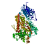
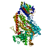
| Title | Soybean Lipoxygenase (LOX-3) at ambient temperatures at 2.0 A resolution | ||||||
 Components Components | Seed lipoxygenase-3 | ||||||
 Keywords Keywords | OXIDOREDUCTASE / iron metalloprotein / lipoxygenase | ||||||
| Function / homology |  Function and homology information Function and homology informationlinoleate 9S-lipoxygenase / linoleate 9S-lipoxygenase activity / oxylipin biosynthetic process / lipid oxidation / oxidoreductase activity, acting on single donors with incorporation of molecular oxygen, incorporation of two atoms of oxygen / fatty acid biosynthetic process / iron ion binding / cytoplasm Similarity search - Function | ||||||
| Biological species |  | ||||||
| Method |  X-RAY DIFFRACTION / MOLECULAR REPLACEMENT / Resolution: 2 Å X-RAY DIFFRACTION / MOLECULAR REPLACEMENT / Resolution: 2 Å | ||||||
 Authors Authors | Borbulevych, O.Y. / Jankun, J. / Skrzypczak-Jankun, E. | ||||||
 Citation Citation | Journal: Acta Crystallogr.,Sect.D / Year: 2006 Title: Effect of crystal freezing and small-molecule binding on internal cavity size in a large protein: X-ray and docking studies of lipoxygenase at ambient and low temperature at 2.0 A resolution. Authors: Skrzypczak-Jankun, E. / BORBULEVYCH, O.Y. / ZAVODSZKY, M.I. / BARANSKI, M.R. / PADMANABHAN, K. / PETRICEK, V. / JANKUN, J. #1: Journal: Proteins / Year: 1997Title: Structure of soybean lipoxygenase L3 and a comparison with its L1 isoenzyme Authors: Skrzypczak-Jankun, E. / Amzel, L.M. / Kroa, B. / Funk, M.O. #2: Journal: Acta Crystallogr.,Sect.D / Year: 1996Title: Flash-freezing causes a stress induced modulation in a crystal structure of soybean lipoxygenase L3 Authors: Skrzypczak-Jankun, E. / Bianchet, M. / Amzel, L.M. / Funk, M.O. | ||||||
| History |
|
- Structure visualization
| Structure viewer | Molecule: MolmilJmol/JSmol |
|---|
- Downloads & links
-Download
| PDBx/mmCIF format | 1rrh.cif.gz | 193.7 KB | Display | PDBx/mmCIF format |
|---|---|---|---|---|
| PDB format | pdb1rrh.ent.gz | 150.9 KB | Display | PDB format |
| PDBx/mmJSON format | 1rrh.json.gz | Tree view | PDBx/mmJSON format | |
| Others |  Other downloads Other downloads |
-Validation report
| Arichive directory | https://data.pdbj.org/pub/pdb/validation_reports/rr/1rrhftp://data.pdbj.org/pub/pdb/validation_reports/rr/1rrh | HTTPS FTP |
|---|
-Related structure data
| Related structure data |  1rrlC  1no3S S: Starting model for refinement C: citing same article ( |
|---|---|
| Similar structure data |


-Links
 PDBj
PDBj


- Assembly
| Deposited unit | 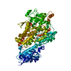
| |||||||||
|---|---|---|---|---|---|---|---|---|---|---|
| 1 |
| |||||||||
| Unit cell |
| |||||||||
| Components on special symmetry positions |
|
-Components
| #1: Protein | Mass: 96919.000 Da / Num. of mol.: 1 / Source method: isolated from a natural source / Source: (natural) |
|---|---|
| #2: Chemical | ChemComp-FE2 /   Mass: 55.845 Da / Num. of mol.: 1 / Source method: obtained synthetically / Formula: Fe Mass: 55.845 Da / Num. of mol.: 1 / Source method: obtained synthetically / Formula: Fe |
| #3: Water | ChemComp-HOH /  Mass: 18.015 Da / Num. of mol.: 549 / Source method: isolated from a natural source / Formula: H2O Mass: 18.015 Da / Num. of mol.: 549 / Source method: isolated from a natural source / Formula: H2O |
| Sequence details | VARIANT RESIDUES IN STRAIN PROVAR ARE NOTED IN SWISS-PROT ENTRY P09186. |
-Experimental details
-Experiment
| Experiment | Method: X-RAY DIFFRACTION / Number of used crystals: 2 |
|---|
- Sample preparation
| Crystal | Density Matthews: 2.45 Å3/Da / Density % sol: 49.84 % |
|---|---|
| Crystal grow | Temperature: 295 K / Method: vapor diffusion, sitting drop / pH: 5.3 Details: 20% PEG 8000, citrate-phosphate buffer 0.05M, tris.HCl, 0.2% sodium azide, pH 5.3, VAPOR DIFFUSION, SITTING DROP, temperature 295K |
-Data collection
| Diffraction | Mean temperature: 295 K |
|---|---|
| Diffraction source | Source: ROTATING ANODE / Type: RIGAKU RU200 / Wavelength: 1.5418 Å |
| Detector | Type: RIGAKU RAXIS II / Detector: IMAGE PLATE / Date: Nov 15, 1997 / Details: graphite monochromator |
| Radiation | Monochromator: GRAPHITE / Protocol: SINGLE WAVELENGTH / Monochromatic (M) / Laue (L): M / Scattering type: x-ray |
| Radiation wavelength | Wavelength: 1.5418 Å / Relative weight: 1 |
| Reflection | Resolution: 2→99 Å / Num. all: 62944 / Num. obs: 54258 / % possible obs: 86.2 % / Observed criterion σ(F): 1 / Observed criterion σ(I): 0 / Redundancy: 2 % / Biso Wilson estimate: 40.6 Å2 / Rmerge(I) obs: 0.079 / Net I/σ(I): 13.5 |
| Reflection shell | Resolution: 2→2.07 Å / Redundancy: 1 % / Rmerge(I) obs: 0.445 / Mean I/σ(I) obs: 1.47 / Num. unique all: 4817 / % possible all: 0.767 |
- Processing
| Software |
| ||||||||||||||||||||||||||||||||||||||||||||||||||||||||||||||||||||||||||||||||||||||||||||||||||||||||||||||||||||||||||||||||||||||||||||||||||||||||||||||||
|---|---|---|---|---|---|---|---|---|---|---|---|---|---|---|---|---|---|---|---|---|---|---|---|---|---|---|---|---|---|---|---|---|---|---|---|---|---|---|---|---|---|---|---|---|---|---|---|---|---|---|---|---|---|---|---|---|---|---|---|---|---|---|---|---|---|---|---|---|---|---|---|---|---|---|---|---|---|---|---|---|---|---|---|---|---|---|---|---|---|---|---|---|---|---|---|---|---|---|---|---|---|---|---|---|---|---|---|---|---|---|---|---|---|---|---|---|---|---|---|---|---|---|---|---|---|---|---|---|---|---|---|---|---|---|---|---|---|---|---|---|---|---|---|---|---|---|---|---|---|---|---|---|---|---|---|---|---|---|---|---|---|
| Refinement | Method to determine structure: MOLECULAR REPLACEMENT Starting model: 1NO3 Resolution: 2→10 Å / Cor.coef. Fo:Fc: 0.959 / Cor.coef. Fo:Fc free: 0.934 / SU B: 4.833 / SU ML: 0.133 / TLS residual ADP flag: LIKELY RESIDUAL / Cross valid method: THROUGHOUT / σ(F): 0 / ESU R: 0.221 / ESU R Free: 0.186 / Stereochemistry target values: Engh & Huber / Details: MAXIMUM LIKELIHOOD refinement
| ||||||||||||||||||||||||||||||||||||||||||||||||||||||||||||||||||||||||||||||||||||||||||||||||||||||||||||||||||||||||||||||||||||||||||||||||||||||||||||||||
| Solvent computation | Ion probe radii: 0.8 Å / Shrinkage radii: 0.8 Å / VDW probe radii: 1.4 Å / Solvent model: BABINET MODEL WITH MASK | ||||||||||||||||||||||||||||||||||||||||||||||||||||||||||||||||||||||||||||||||||||||||||||||||||||||||||||||||||||||||||||||||||||||||||||||||||||||||||||||||
| Displacement parameters | Biso mean: 25.957 Å2
| ||||||||||||||||||||||||||||||||||||||||||||||||||||||||||||||||||||||||||||||||||||||||||||||||||||||||||||||||||||||||||||||||||||||||||||||||||||||||||||||||
| Refinement step | Cycle: LAST / Resolution: 2→10 Å
| ||||||||||||||||||||||||||||||||||||||||||||||||||||||||||||||||||||||||||||||||||||||||||||||||||||||||||||||||||||||||||||||||||||||||||||||||||||||||||||||||
| Refine LS restraints |
| ||||||||||||||||||||||||||||||||||||||||||||||||||||||||||||||||||||||||||||||||||||||||||||||||||||||||||||||||||||||||||||||||||||||||||||||||||||||||||||||||
| LS refinement shell | Resolution: 2→2.05 Å / Total num. of bins used: 20 /
| ||||||||||||||||||||||||||||||||||||||||||||||||||||||||||||||||||||||||||||||||||||||||||||||||||||||||||||||||||||||||||||||||||||||||||||||||||||||||||||||||
| Refinement TLS params. | Method: refined / Origin x: 27.5134 Å / Origin y: 4.2078 Å / Origin z: 15.1744 Å
|
