 Movie
Movie Controller

[English] 日本語
 Yorodumi
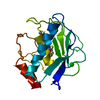
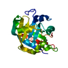
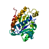
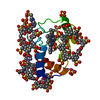
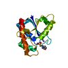
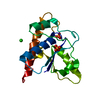
- PDB-1rm8: Crystal structure of the catalytic domain of MMP-16/MT3-MMP: Char... -
+ Open data

- Basic information
| Entry | Database: PDB / ID: 1rm8 | ||||||
|---|---|---|---|---|---|---|---|
| Title | Crystal structure of the catalytic domain of MMP-16/MT3-MMP: Characterization of MT-MMP specific features | ||||||
 Components Components | Matrix metalloproteinase-16 | ||||||
 Keywords Keywords | HYDROLASE / MMP-16 / MT3-MMP / MT-MMP / Membrane Type - Matrix Metalloproteinase / Batimastat / Hydroxamate inhibitor / Protease | ||||||
| Function / homology |  Function and homology information Function and homology informationTGFBR3 PTM regulation / Hydrolases; Acting on peptide bonds (peptidases); Metalloendopeptidases / Activation of Matrix Metalloproteinases / metalloaminopeptidase activity / collagen catabolic process / extracellular matrix organization / skeletal system development / protein processing / metalloendopeptidase activity / Golgi lumen ...TGFBR3 PTM regulation / Hydrolases; Acting on peptide bonds (peptidases); Metalloendopeptidases / Activation of Matrix Metalloproteinases / metalloaminopeptidase activity / collagen catabolic process / extracellular matrix organization / skeletal system development / protein processing / metalloendopeptidase activity / Golgi lumen / enzyme activator activity / extracellular matrix / cell surface / proteolysis / : / zinc ion binding / plasma membrane Similarity search - Function | ||||||
| Biological species |  Homo sapiens (human) Homo sapiens (human) | ||||||
| Method |  X-RAY DIFFRACTION / SYNCHROTRON / MOLECULAR REPLACEMENT / Resolution: 1.8 Å X-RAY DIFFRACTION / SYNCHROTRON / MOLECULAR REPLACEMENT / Resolution: 1.8 Å | ||||||
 Authors Authors | Lang, R. / Braun, M. / Sounni, N.E. / Noel, A. / Frankenne, F. / Foidart, J.-M. / Bode, W. / Maskos, K. | ||||||
 Citation Citation | Journal: J.Mol.Biol. / Year: 2004 Title: Crystal structure of the catalytic domain of MMP-16/MT3-MMP: characterization of MT-MMP specific features. Authors: Lang, R. / Braun, M. / Sounni, N.E. / Noel, A. / Frankenne, F. / Foidart, J.M. / Bode, W. / Maskos, K. | ||||||
| History |
|
- Structure visualization
| Structure viewer | Molecule: MolmilJmol/JSmol |
|---|
- Downloads & links
-Download
| PDBx/mmCIF format | 1rm8.cif.gz | 54.7 KB | Display | PDBx/mmCIF format |
|---|---|---|---|---|
| PDB format | pdb1rm8.ent.gz | 37.1 KB | Display | PDB format |
| PDBx/mmJSON format | 1rm8.json.gz | Tree view | PDBx/mmJSON format | |
| Others |  Other downloads Other downloads |
-Validation report
| Arichive directory | https://data.pdbj.org/pub/pdb/validation_reports/rm/1rm8ftp://data.pdbj.org/pub/pdb/validation_reports/rm/1rm8 | HTTPS FTP |
|---|
-Related structure data
| Related structure data |  1bqqS S: Starting model for refinement |
|---|---|
| Similar structure data |


-Links
 PDBj
PDBj


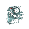
- Assembly
| Deposited unit | 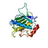
| ||||||||||
|---|---|---|---|---|---|---|---|---|---|---|---|
| 1 |
| ||||||||||
| Unit cell |
|
-Components
| #1: Protein | Mass: 18874.764 Da / Num. of mol.: 1 / Fragment: catalytic domain Source method: isolated from a genetically manipulated source Details: complexed with Batimastat (bb94) / Source: (gene. exp.) Homo sapiens (human) / Plasmid: pT7cdMP3 / Species (production host): Escherichia coli / Production host:  References: UniProt: P51512, Hydrolases; Acting on peptide bonds (peptidases); Metalloendopeptidases | ||||||
|---|---|---|---|---|---|---|---|
| #2: Chemical |   Mass: 65.409 Da / Num. of mol.: 2 / Source method: obtained synthetically / Formula: Zn Mass: 65.409 Da / Num. of mol.: 2 / Source method: obtained synthetically / Formula: Zn#3: Chemical |   Mass: 40.078 Da / Num. of mol.: 2 / Source method: obtained synthetically / Formula: Ca Mass: 40.078 Da / Num. of mol.: 2 / Source method: obtained synthetically / Formula: Ca#4: Chemical | ChemComp-BAT / | 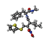  Mass: 477.640 Da / Num. of mol.: 1 / Source method: obtained synthetically / Formula: C23H31N3O4S2 / Comment: inhibitor*YM Mass: 477.640 Da / Num. of mol.: 1 / Source method: obtained synthetically / Formula: C23H31N3O4S2 / Comment: inhibitor*YM#5: Water | ChemComp-HOH / |  Mass: 18.015 Da / Num. of mol.: 115 / Source method: isolated from a natural source / Formula: H2O Mass: 18.015 Da / Num. of mol.: 115 / Source method: isolated from a natural source / Formula: H2O |
-Experimental details
-Experiment
| Experiment | Method: X-RAY DIFFRACTION / Number of used crystals: 2 |
|---|
- Sample preparation
| Crystal | Density Matthews: 2.59 Å3/Da / Density % sol: 52.55 % | ||||||||||||||||||||||||||||||||||||||||||||||||||||||||
|---|---|---|---|---|---|---|---|---|---|---|---|---|---|---|---|---|---|---|---|---|---|---|---|---|---|---|---|---|---|---|---|---|---|---|---|---|---|---|---|---|---|---|---|---|---|---|---|---|---|---|---|---|---|---|---|---|---|
| Crystal grow | Temperature: 293 K / Method: vapor diffusion, sitting drop / pH: 8.5 Details: Tris-HCl, Sodium/potassium tartrate, pH 8.5, VAPOR DIFFUSION, SITTING DROP, temperature 293K | ||||||||||||||||||||||||||||||||||||||||||||||||||||||||
| Crystal grow | *PLUS Method: vapor diffusion, sitting drop | ||||||||||||||||||||||||||||||||||||||||||||||||||||||||
| Components of the solutions | *PLUS
|
-Data collection
| Diffraction |
| ||||||||||||||||||
|---|---|---|---|---|---|---|---|---|---|---|---|---|---|---|---|---|---|---|---|
| Diffraction source |
| ||||||||||||||||||
| Detector |
| ||||||||||||||||||
| Radiation | Monochromator: GRAPHITE / Protocol: SINGLE WAVELENGTH / Monochromatic (M) / Laue (L): M / Scattering type: x-ray | ||||||||||||||||||
| Radiation wavelength |
| ||||||||||||||||||
| Reflection | Resolution: 1.8→16.6 Å / Num. obs: 18455 / % possible obs: 96.7 % / Observed criterion σ(I): 2.2 / Biso Wilson estimate: 23.51 Å2 / Rsym value: 0.083 / Net I/σ(I): 5.5 | ||||||||||||||||||
| Reflection shell | Resolution: 1.8→1.9 Å / Rmerge(I) obs: 0.327 / Mean I/σ(I) obs: 2.2 / Num. unique all: 1090 / Rsym value: 0.327 / % possible all: 91.1 | ||||||||||||||||||
| Reflection | *PLUS Lowest resolution: 16.6 Å / Num. measured all: 119029 / Rmerge(I) obs: 0.083 | ||||||||||||||||||
| Reflection shell | *PLUS Highest resolution: 1.8 Å / Lowest resolution: 1.9 Å / % possible obs: 91.1 % |

- Processing
| Software |
| |||||||||||||||||||||||||
|---|---|---|---|---|---|---|---|---|---|---|---|---|---|---|---|---|---|---|---|---|---|---|---|---|---|---|
| Refinement | Method to determine structure: MOLECULAR REPLACEMENT Starting model: Pdb code 1bqq Resolution: 1.8→16.6 Å / Isotropic thermal model: overall temperature factors / Cross valid method: FREE R-VALUE / σ(F): 1 / Stereochemistry target values: Engh & Huber
| |||||||||||||||||||||||||
| Displacement parameters | Biso mean: 23.509 Å2
| |||||||||||||||||||||||||
| Refinement step | Cycle: LAST / Resolution: 1.8→16.6 Å
| |||||||||||||||||||||||||
| Refine LS restraints |
| |||||||||||||||||||||||||
| Software | *PLUS Version: 5 / Classification: refinement | |||||||||||||||||||||||||
| Refinement | *PLUS Lowest resolution: 16.6 Å / Num. reflection obs: 17555 / % reflection Rfree: 4.9 % / Rfactor Rfree: 0.236 / Rfactor Rwork: 0.208 | |||||||||||||||||||||||||
| Solvent computation | *PLUS | |||||||||||||||||||||||||
| Displacement parameters | *PLUS | |||||||||||||||||||||||||
| Refine LS restraints | *PLUS
|
